
Ameliorative Effect of Aster scaber Thunberg and Chaenoleles sinensis Koehne Complex Extracts Against Oxidative Stress-induced Memory Dysfunction in PC12 Cells and ICR Mice
This is an open access article distributed under the terms of the Creative Commons Attribution Non-Commercial License (http://creativecommons.org/licenses/by-nc/3.0/) which permits unrestricted non-commercial use, distribution, and reproduction in any medium, provided the original work is properly cited.
Abstract
Oxidative stress plays an important role in neuro-degenerative disorders such as Alzheimer's disease. Oxidative stress is mediated by reactive oxygen species (ROS), which are implicated in the pathogenesis of numerous diseases, and account for the toxicity of a wide range of compounds.
In order to study the neuro-protective effect of the complex extracts of Aster scaber Thunberg (AS) and Chaenoleles sinensis Koehne (CSK) against hydrogen peroxide in PC12 cells, cell viability was evaluated by the MTT assay using tetrazole, 3-(4,5-dimethylthiazol-2-yl)-2,5-diphenyltetrazolium bromide and the intracellular ROS levels were determined the by 2’,7’-dichlorofluorescein diacetate (DCF-DA) assay. In order to examine the anti-amnesic effects of the complex extracts of AS and CSK, behavioral tests were performed on male ICR mice. The ameliorating effect of the complex extracts against Aβ1-42-induced learning and memory impairment was analyzed by y-maze and passive avoidance tests. The AS and CSK extracts showed neuro-protective activity both in vitro and in vivo, and the neuro-protective effect of their 60 : 40 (AS : CSK) mixture was better than that of the other mixtures. Moreover, the complex extracts synergistically inhibited acetylcholinesterase activity and rapid peroxidation.
A mixture of the AS and CSK extracts could be used to develop functional foods and serve as raw materials for the development of therapeutics against Alzheimer's disease.
Keywords:
Aster scaber Thunberg (AS), Chaenoleles sinensis Koehne (CSK), Complex Extracts, Oxidative Stress, Reactive Oxygen Species, Synergy EffectINTRODUCTION
One of the characteristics of Alzheimer's disease (AD) is increased levels of intracellular reactive oxygen species (ROS), resulting from the accumulation of amyloid beta peptide (Aβ). Aβ is produced through the amyloidogenic pathway, via sequential cleavage of amyloid precursor protein (APP) by β- and γ-secretases (Choi et al., 2013). Accumulation of this peptide could be the main pathogenic mechanism of AD and may trigger neurotoxicity (Jin et al., 2009; Xiaowei et al., 2015).
ROS are considered to be the main cause of a wide range of common diseases, including cardiovascular diseases, inflammatory conditions, cancer, mutations, and neuro-degenerative diseases (George et al., 2002). Increased ROS levels lead to oxidative stress and modify the intracellular components, causing DNA breakage, lipid peroxidation, and protein carbonylation (George et al., 2002; Cheignon et al., 2018). This induces cytotoxicity and fatal cellular damage.
Antioxidants are vital substances that protect the body from ROS-mediated damage. Plants have complex antioxidant systems to inhibit the oxidative chain reaction. They also produce phytochemicals, which are bioactive compounds obtained from secondary metabolites (Sridhar and Charles, 2019). Some of these phytochemicals exhibit high antioxidant activity and exert other possible effects to decrease the risk of neuro-degenerative diseases caused by Aβ-induced oxidative stress (Jeong et al., 2010; Sridhar and Charles, 2019).
Aster scaber Thunberg (AS) is a perennial plant belonging to the chrysanthemum family and grows in mountainous areas and grasslands of Asia, including Korea. It is rich in various nutrients, including calcium, iron, flavonoids, and saponin. It is reported that the anti-cancer and anti-inflammatory activity of these constituent in AS extracts. Also, another study confirmed acetylcholinesterase inhibition activity of AS extracts according to different extraction processes (Kim and Youn, 2014).
Chaenoleles sinensis Koehne (CSK) belongs to the Rosaceae family found in the East Asia region, including Korea. It is rich in dietary fibers, organic acids and bioactive triterpenes, such as oleanolic acid and ursolic acid. It also contains large amounts of bioactive phenolic acids and vitamins. In particular, the flavonoids of CSK are the main phytochemical constituents that have proven effective in preventing oxidative damage caused by free radicals (Zhang et al., 2009). According to Sancheti and Seo’s study, ethanol extracts of CSK showed antiacetylcholinesterase effects in diabetic rats (Sancheti et al., 2013).
To develop preventive strategies for neuro-degenerative diseases like AD, we investigated the antioxidative and neuroprotective effects of AS and CSK complex extracts against H2O2-induced cytotoxicity in PC12 cells. We also investigated the cognitive-enhancing effect of AS and CSK complex extracts on Aβ-induced learning and memory impairment in ICR (Institute of Cancer Research) mice.
MATERIALS AND METHODS
1. Chemicals
Dimethyl sulfoxide (DMSO), hydrogen peroxide (H2O2), L-ascorbic acid (vitamin C), 3-(4,5-dimethylthiazol-2-yl)-2,5-diphenyltetrazolium bromide (MTT), 2',7'-dichlorofluorescein diacetate (DCF-DA), 2-thiobarbituric acid (TBA), amyloid beta peptide (Aβ1-42, Aβ42-1) were purchased from Sigma-Aldrich (St. Louis, MO, USA). 2,2'-azobis-(2-amidinopropane) dihydrochloride (AAPH) was purchased from Wako Pure Chemical (Richmond, VA, USA). All other chemicals used were of analytical grade.
2. Sample preparation
Aster scaber Thunberg and Chaenoleles sinensis Koehne were obtained from a local market (Sancheong and Cheongdo, Korea). After drying, all the samples were ground to powder and extracted with 10 volumes of water at 90℃ for 4 h respectively. The extracts were filtered through a Whatman filter paper No.1 (Whatman Co., Maidstone, England) and dried in a rotary evaporator (EYELA, Tokyo Rikakikai Co., Ltd., Tokyo, Japan) under reduced pressure at 37℃. As a result, the dried sample AS (6.4 ㎏) and CSK (3 ㎏) were extracted using hot water extraction, the percentage yield of these extracts were 14.5% and 29.6% w/w, respectively. And then, AS and CSK extracts were prepared at eleven different concentrations (AS : CSK = 100 : 0, 90 : 10, 80 : 20, 70 : 30, 60 : 40, 50 : 50, 40 : 60, 30 : 70, 20 : 80, 10 : 90, 0 : 100, w/w).
3. DPPH radical scavenging activity assay
2,2-diphenyl-1-picrylhydrazyl (DPPH) assay was carried out according to Blois method with a slight modification (Blois, 1958). Briefly, a 0.2 mM solution of DPPH radical solution in ethanol was prepared, and then, 1.5 mL of this solution was mixed with 0.5 ㎖ of the extract solution; finally, after 30 min, the absorbance was measured at 517 ㎚ (SpectraMax M2, Molecular Devices Llc., Sunnyvale, CA, USA). Vitamin C was used as the positive control. The scavenging activity is shown as percentage DPPH scavenging, calculated as
4. Cell culture
The PC12 cell line (CRL-1721) was purchased from the American Type Culture Collection (ATCC, Manassas, VA, USA). The PC12 cells were cultured in RPMI 1640 medium supplemented with 10% (v/v) horse serum, 5% (v/v) fetal bovine serum, and 1% (v/v) antibiotic-antimycotic (Gibco BRL Co., Gaithersburg, MD, USA). Cells were cultured in 100 ㎜ tissue culture dishes (Falcon Inc., NY, USA). Cultures were maintained at 37℃ with 5% CO2 and water saturation.
5. Assessment of cell viability
Cell viability was determined by MTT [3-(4,5-dimethylthiazol-2-yl)-2,5-diphenyltetrazolium bromide] tetrazolium reduction assay, wherein the yellow tetrazole is reduced to a purple insoluble compound, known as formazan, in the mitochondria of metabolically active cells. Cells were seeded in 96-well plates at a density of 105 - 106 cells/㎖ (Choi et al., 2009).
In the cytotoxicity studies, PC12 cells were treated with H2O2 at the indicated concentration for 2 h at 37℃. Finally, cells were incubated for 3 h at 37℃ with MTT (2.5 ㎎/㎖ final concentration). Then, the formazan crystals were dissolved in DMSO. The absorbance was measured on a microplate reader (SpectraMax M2, Molecular Devices Llc., Sunnyvale, CA, USA) using a reference wavelength of 630 ㎚ and a test wavelength of 570 ㎚.
6. Measurement of oxidative stress
Levels of cellular oxidative stress were measured using DCF-DA. PC12 cells were pretreated for 12 h with Aster scaber Thunberg and Chaenoleles sinensis Koehne complex extracts. Cells were then treated with or without H2O2 for 2 h. At the end of the treatment, cells were incubated in the presence of 50 μM DCF-DA for 50 min (Choi et al., 2009). After incubation, DCF was quantified using the SpectraMax M2 (Molecular Devices Llc., Sunnyvale, CA, USA). The results were expressed as percent relative to the oxidative stress level of the control cells, which was set to 100%.
7. Animals
The ICR mice (males, 5-week-old) were purchased from Daehan Biolink (Chungnam, Korea). Eight mice were housed per cage in a room maintained at 24 ± 1℃ and 55% humidity with an alternating 12 h light-dark cycle. They had free access to feed and water. All experiments were conducted according to the guidelines of the Committee on Care and Use of Laboratory Animals of Korea University (certificate: kuiacuc-2018-75).
Subsequently, Aβ1-42 was administered via intracerebroventricular (ICV) injection. The control group was injected with Aβ42-1. Briefly, Aβ was dissolved in 0.85% sodium chloride solution (v/v). Each mouse was injected at the bregma with a 25 ㎕ Hamilton microsyringe fitted with a 26-gauge needle that was inserted to a depth of 2.5 ㎜. The injection volume was 5 ㎕ (dose 410 p㏖/mouse) (Yan et al., 2001). The sample groups were injected with Aβ1-42 after their diets were supplemented with AS and CSK extract mixture (AS 400; AS : CSK = 100 : 0, 400 ㎎/㎏, CSK 400; AS : CSK = 0 : 100, 400 ㎎/㎏, AS-CSK 400; AS : CSK = 60 : 40, 400 ㎎/㎏, AS-CSK 800; AS : CSK = 60 : 40, 800 ㎎/㎏).
8. Y-maze test
The Y-maze test was carried out 3 days after the Aβ injection. The maze was made of black opaque Y-shaped plastic, and each arm of the maze was 33 ㎝ long, 15 ㎝ high, 10 ㎝ wide, and positioned at an equal angle. Each mouse was placed at the end of one arm and allowed to move freely through the maze during an 8 min period. The sequence of the arm entries was recorded manually.
A spontaneous alternation behavior was defined as entry into all three arms on consecutive choices in overlapping triplet sets. The percentage of spontaneous alternation behavior was calculated as the ratio of actual to possible alternations (Zhang et al., 2009; Choi et al., 2017).
9. Passive avoidance test
After the Y-maze test, the passive avoidance test was conducted to examine short-term memory. The apparatus consisted of a light room and a dark room. The dark room had a grid of steel rods connected to a shock generator. The rooms were separated by a wall with a connecting passage. For the acquisition trial, each mouse was put into the light room and allowed to explore freely (for 1 min, with no light or shock). Each mouse was allowed to explore further for 2 min with light and no shock.
A mouse that had completely entered into the dark room received a foot shock of 0.5 ㎃ for 1s through the steel grid. One day later, the same mouse was again put into the light room, and the latency time taken to enter the dark room was measured without any foot shock. The maximum cut-off latency was set to 300 s. Doses of samples were designed 400 ㎎/㎏ and 800 ㎎/㎏ according to previous study (Kwon et al., 2016; Choi et al., 2017).
10. Determination of lipid peroxidation in mouse brain tissue
The brains were homogenized in cold PBS in an ice bath. The homogenates were directly centrifuged twice at a 30 s interval at 10,000 rpm for 10 s. Aliquots of the supernatant were used to determine the malondialdehyde (MDA) levels and protein content in the brain.
The MDA level was assayed for lipid peroxidation products by using a previously described method (Blois, 1958). Briefly, 80 ㎕ of each homogenate was mixed with 480 ㎕ phosphoric acid (1%, v/v), followed by the addition of 160 ㎕ thiobarbituric acid solution (0.67%, w/v). The mixture was incubated at 95℃ in a water bath for 45 min. After cooling, the colored complex was extracted with n-butanol. The butanol phase was separated by centrifugation, and the absorbance was measured using tetramethoxypropane as a standard at 532 ㎚.
11. Measurement of acetylcholinesterase (AChE) activity in mouse brain tissue
AChE activity was measured by Ellman's modified method. Briefly, PC12 cells were homogenized with Tris-HCl buffer (20 mM Tris-HCl, pH 7.5 containing 150 mM NaCl, 10 mM MgCl2, and 0.5% Triton X-100), and then, the samples were centrifuged at 10,000 × g for 15 min. The supernatant was used as an enzyme source, and acetylcholine iodide (ACh iodine) was used as the reaction substrate. 5,5'-dithiobis-2-nitrobenzoic acid (DTNB) was used to quantify the thiocholine produced from the hydrolysis of AChE.
A 10 ㎕ liquot of each sample was mixed with 10 ㎕ of enzyme solution, which was subsequently added to 70 ㎕ of the reaction mixture (50 mM sodium phosphate buffer, pH 8.0, containing 1 mM DTNB and 0.5 mM ACh iodine). This mix was then incubated at 37℃ for 15 min (Ellman et al., 1961). The final enzyme reaction was monitored at a wavelength of 405 ㎚ using a microplate reader (SpectraMax M2, Molecular Devices Llc., Sunnyvale, CA, USA). The percentage of inhibition was calculated by comparing the rates of absorbance.
12. Quantification of ACh content in mouse brain tissue
The ACh content in the brain was measured using the method of Hestrin, as described previously, based on the reaction of ACh and hydroxylamine. Mice were sacrificed, and their brains maintained at -80℃, prior to use. The brains were homogenized in cold phosphate buffered saline (PBS) in an ice bath. The homogenates were directly centrifuged twice at a 30 s interval at 10,000 rpm for 10 s.
Subsequently, 1 ㎖ of the homogenate was mixed with 2 ㎖ alkaline hydroxylamine reagent. After at least 1min, the pH was adjusted to 1.2 ± 0.2 with 1 ㎖ HCl solution and 1 ml iron solution. The density of the purple brown color was determined at 540 ㎚ (Hestrin, 1949).
13. Statistical analysis
All results were presented as means ± standard deviation (SD). Statistical analyses were performed using the Statistical Analysis System (SAS 9.4) software package (Cary, NC, USA).
Significant differences between groups were examined by one-way ANOVA followed by the Scheffe's Multiple Range Test. A p-value < 0.05 was considered statistically significant.
RESULTS
1. DPPH radical scavenging activity
The DPPH radical scavenging activities of the AS and CSK extracts were estimated through comparing the percentage inhibition of the formation of DPPH radicals by the concentrations of AS, CSK, and vitamin C. The reduction capability of DPPH was determined by the decrease in its absorbance at 517 ㎚, which is induced by antioxidants. A 1 ㎎/㎖ of AS, CSK, and vitamin C exhibited 77.7, 71.3, and 133.3% inhibition, respectively (Fig. 1). The RC50 values of AS, CSK, and vitamin C for DPPH radical scavenging activity were 0.207 ㎎/㎖, 0.168 ㎎/㎖, 0.016 ㎎/㎖, respectively.
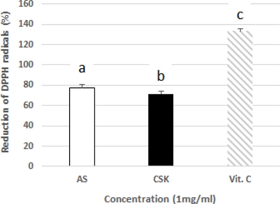
DPPH radical scavenging activities of AS and CSK extracts.Vit. C group; L-ascorbic acid (1 ㎎/㎖), the sample groups (□; AS 1 ㎎/㎖, ■; CSK, 1 ㎎/㎖). Each value represents the means ± SD (n = 3). Between groups comparisons were conducted using One-way ANOVA with Scheffe's Multiple Range Test (Different superscripts indicate significant difference among groups at p < 0.05).
2. Cell viability
To confirm the synergistic effect of AS and CSK complex extracts on the cell viability, MTT reduction assay was performed. The yellow tetrazole is reduced to a purple insoluble compound, known as formazan, in the mitochondria of metabolically active cells. As shown in Fig. 2, the H2O2 group represented low cell viability (35% decrease) compared to the control group (100%). In the sample groups, the mixture was tested at various ratios, and the extract mixture groups (AS : CSK = 60 : 40 – 40 : 60) showed high cell viability against H2O2-induced oxidative stress.
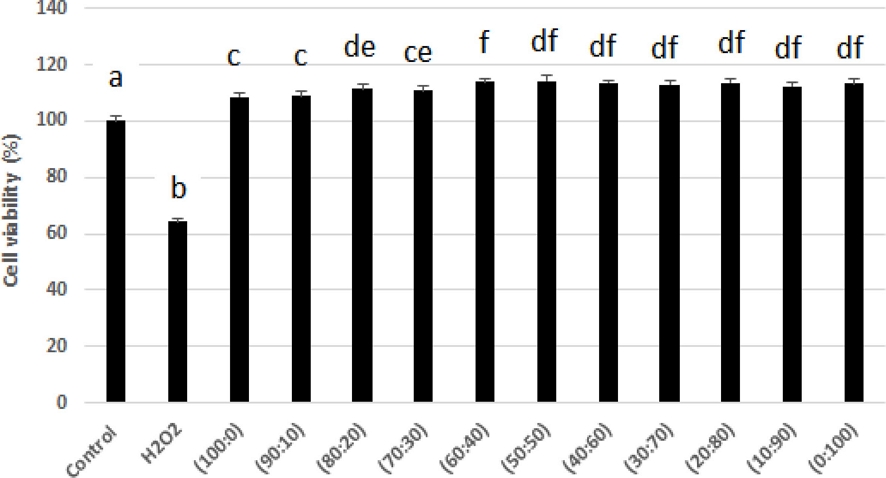
Protective effect of AS and CSK complex extracts against H2O2-induced cytotoxicity.Control group; untreated, H2O2 group; 200 ?M H2O2, Sample group; treatment with various mixtures of AS and CSK complex extracts (100 : 0, 90 : 10, 80 : 20, 70 : 30, 60 : 40, 50 : 50, 40 : 60, 30 : 70, 20 : 80, 10 : 90, 0 : 100, w/w). The concentration of all the samples was 1 ㎎/㎖. Each value represents the means ± SD (n = 4). *Mean within a column followed by the same letters are not significantly different based on One-way ANOVA with Scheffe's Multiple Range Test (Different superscripts indicate significant difference among groups at p < 0.05).
3. Oxidative stress
To measure the neuro-protective effects of the AS and CSK extract mixtures on H2O2-induced oxidative stress, we evaluated ROS production levels using DCF-DA. The DCF-DA assay was performed to confirm the reduction of H2O2-induced intracellular ROS production in PC12 cells.
The H2O2 group displayed a significant increase in intracellular ROS production (1,860%) compared with the control group (100%). Specifically, the 60 : 40 group exhibited remarkable intracellular ROS reduction (384%) (Fig. 3).
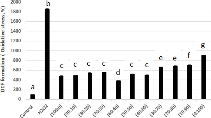
Protective effect of AS and CSK complex extracts against oxidative stress in PC12.Control group; untreated, H2O2 group; 200 mM H2O2, Sample group; treatment with various mixtures of AS and CSK complex extracts (100 : 0, 90 : 10, 80 : 20, 70 : 30, 60 : 40, 50 : 50, 40 : 60, 30 : 70, 20 : 80, 10 : 90, 0 : 100, w/w). ㎎/㎖. Each value represents the means ± SD (n = 4). *Mean within a column followed by the same letters are not significantly different based on One-way ANOVA with Scheffe's Multiple Range Test (Different superscripts indicate significant difference among groups at p < 0.05).
4. Y maze test results
To confirm the ameliorating effect of AS and CSK extract mixture on Aβ1-42-induced learning- and memory-impaired mice, the Y-maze test was conducted. The Y-maze results displayed impairment of the spatial cognitive function of the Aβ group (58.16%) compared to the alternation behavior of the control group (73.20%). The AS and CSK extract diet group showed slightly increased alternation behavior (AS; 63.41%, CSK; 61.87%) (Fig. 4A).

Memory ameliorating effects of AS and CSK complex extracts against Aβ1-42 induced cognitive impairment in the Y-maze test.The control group was injected with Aβ42-1. The Aβ group was injected with 410 pmol of Aβ1-42 per mouse. The sample groups (AS400, CSK400, AS-CSK 400, AS-CSK 800) were injected with Aβ1-42 followed by feeding with AS and CSK complex extracts (AS400; AS : CSK = 100 : 0, 400 ㎎/㎏, CSK400; AS : CSK = 0 : 100, 400 ㎎/㎏, AS-CSK400; AS : CSK = 60 : 40, 400 ㎎/㎏, AS-CSK 800; AS : CSK = 60 : 40, 800 ㎎/㎏ body weight per day). The spontaneous alternation behaviors were measured during 8 min. Each value represents the means ± SD. (n = 4 - 8). Mean within a column followed by the same letters are not significantly different based on One-way ANOVA with Scheffe's Multiple Range Test (*,#,§,**Different superscripts indicate statistical differences among the groups at p < 0.05).
In contrast, the AS and CSK extract mixture diet group significantly increased spontaneous alternation behavior after Aβ injection (AS-CSK 400 = 68.20%, AS-CSK 800 = 76.695%). The number of arm entries did not change in all the experimental groups (Fig. 4B).
5. Passive avoidance test results
The passive avoidance test reflects short-term learning and memory function. The Aβ1-42 injected mice exhibited a significant reduction (21% decrease) in the step through latency compared to the control group (Fig. 5).
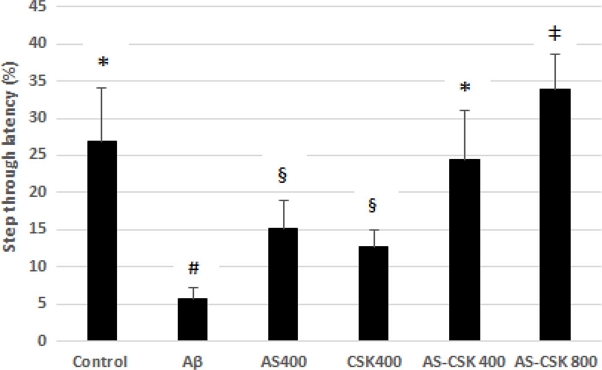
Memory ameliorating effects of AS and CSK complex extracts against Aβ1-42 induced cognitive impairment through passive avoidance test.Control group was injected with Aβ42-1. Aβ group was injected with 410 pmol of Aβ1-42 per mouse. Sample groups (AS400, CSK400, AS-CSK 400, AS-CSK 800) were injected with Aβ1-42 followed by feeding with AS and CSK complex extracts (AS400; AS : CSK = 100 : 0, 400 ㎎/㎏, CSK400; AS : CSK = 0 : 100, 400 ㎎/㎏, AS-CSK 400; AS : CSK = 60 : 40, 400 ㎎/㎏, AS-CSK 800; AS : CSK = 60 : 40, 800 ㎎/㎏ body weight per day). The step-through latency was measured during 5 min. Each value represents the means ± SD. (n = 4 - 8). Mean within a column followed by the same letters are not significantly different based on one-way ANOVA with Scheffe's Multiple Range Test (*,#,§,‡Different superscripts indicate statistical differences among the groups at p < 0.05).
Similar to the Y-maze test, the AS and CSK extract mixture diet group effectively reversed the Aβ1-42 induced cognitive impairment. In particular, the 60 : 40 mixture groups exhibited a dose-dependent (24.3%, 33.8%) attenuation of Aβ-induced memory impairment.
6. Lipid peroxidation
Lipid peroxidation was evaluated by measuring malondialdehyde (MDA) present in the sample. The levels of MDA were significantly increased in the Aβ-injected group (0.718 μ㏖/㎎·protein) when compared with those of the control group (0.436 μ㏖/㎎·protein). In contrast, the AS and CSK extract mixture diet groups (AS-CSK 400 : 0.31 μ㏖/㎎·protein, AS-CSK 800 : 0.247 μ㏖/㎎·protein) significantly decreased the MDA contents in a dose-dependent manner (Fig. 6).
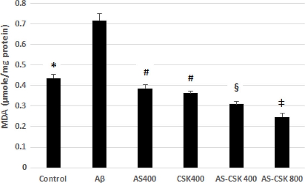
Effect of AS and CSK complex extracts on lipid peroxidation in mice brains. Control group was injected with Aβ42-1.Aβ group was injected with 410 pmol of Aβ1-42 per mouse. Sample groups (AS400, CSK400, AS-CSK 400, AS-CSK 800) were injected with Aβ1-42 followed by feeding with AS and CSK complex extracts (AS400; AS : CSK = 100 : 0, 400 ㎎/㎏, CSK 400; AS : CSK = 0 : 100, 400 ㎎/㎏, AS-CSK400; AS : CSK = 60 : 40, 400 ㎎/㎏, AS-CSK 800; AS : CSK = 60 : 40, 800 ㎎/㎏ body weight per day). Each value represents the means ± SD. (n = 4 - 8). Mean within a column followed by the same letters are not significantly different based on One-way ANOVA with Scheffe's Multiple Range Test (*,#,§,‡Different superscripts indicate statistical differences among the groups at p < 0.05).
7. AChE activity
The Aβ group in this experiment showed an increase (32.8%) in AChE activity compared with the control group. Treatment with extract mixture mitigated the Aβ-induced impairment of the cholinergic system by decreasing AChE activity (Fig. 7).
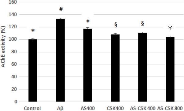
Effect of AS and CSK complex extracts on AChE activity in mice brains. Control group was injected with Aβ42-1.Aβ group was injected with 410 pmol of Aβ1-42 per mouse. Sample groups (AS400, CSK400, AS-CSK 400, AS-CSK 800) were injected with Aβ1-42 followed by feeding with AS and CSK complex extracts (AS400; AS : CSK = 100 : 0, 400 ㎎/㎏, CSK400; AS : CSK = 0 : 100, 400 ㎎/㎏, AS-CSK 400; AS : CSK = 60 : 40, 400 ㎎/㎏, AS-CSK 800; AS : CSK = 60 : 40, 800 ㎎/㎏ body weight per day). Mean within a column followed by the same letters are not significantly different based on one-way ANOVA with Scheffe's Multiple Range Test (*,#,§,‡,¥ Different superscripts indicate statistical differences among the groups at p < 0.05).
8. ACh content
The ACh content in the mouse brain tissue was determined by spectrophotometric analysis. As shown in Fig. 8, the ACh level was lower (39.29 ㎎/㎎·protein) in Aβ-treated mice than in the control group (102.48 ㎎/㎎·protein). In contrast, administration of the AS and CSK extract mixture reversed the decrease in ACh content compared to that of mice treated with Aβ only (AS-CSK 400: 96.69 ㎎/㎎·protein, AS-CSK 800: 94.95 ㎎/㎎·protein).
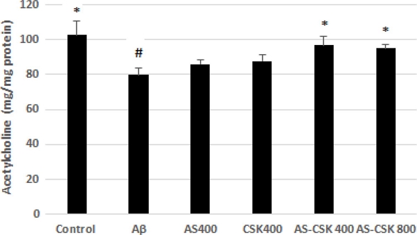
Effect of AS and CSK complex extracts on ACh contents in mice brains.The control group was injected with Aβ42-1. The Aβ group was injected with 410 pmol of Aβ1-42 per mouse. The sample groups (AS400, CSK400, AS-CSK 400, AS-CSK 800) were injected with Aβ1-42 followed by feeding with AS and CSK complex extracts (AS400; AS : CSK = 100 : 0, 400 ㎎/㎏, CSK400; AS : CSK = 0 : 100, 400 ㎎/㎏, AS-CSK 400; AS : CSK = 60 : 40, 400 ㎎/㎏, AS-CSK 800; AS : CSK = 60 : 40, 800 ㎎/㎏ body weight per day). Mean within a column followed by the same letters are not significantly different based on One-way ANOVA with Scheffe's Multiple Range Test (*p < 0.05 as compared with the Aβ group, #p < 0.05 as compared with the control group).
9. AST, ALT activity
After behavioral test, serum was investigated by using a serum transaminase reagent kit. The results of serum transaminases did not represent significant differences among different groups (Fig. 9). It was normal levels of aspartate aminotransferase (AST) and alanine aminotransferase (ALT) ranges for all groups.

Aspartate aminotransferase activity in the serum of ICR mice (A). Alanine aminotransferase activity in the serum of ICR mice (B). The control group was injected with Aβ42-1. The Aβ group was injected with 410 pmol of Aβ1-42 per mouse. The sample groups (AS400, CSK400, AS-CSK 400, AS-CSK 800) were injected with Aβ1-42 followed by feeding with AS and CSK complex extracts (AS400; AS : CSK = 100 : 0, 400 ㎎/㎏, CSK 400; AS : CSK = 0 : 100, 400 ㎎/㎏, AS-CSK 400; AS : CSK = 60 : 40, 400 ㎎/㎏, AS-CSK 800; AS : CSK = 60 : 40, 800 ㎎/㎏ body weight per day). Each value represents the means ± SD. (n = 4 - 8). Mean within a column followed by the same letters are not significantly different based on One-way ANOVA with Scheffe's Multiple Range Test (Different superscripts indicate statistical differences among the groups at p < 0.05).
DISCUSSION
AD is one of the most common forms of dementia affecting approximately 10% of the population over the age of 65 years (Hestrin, 1949). However, the mechanism of Aβ-induced neurotoxicity is still poorly understood (Alzheimer’s Association, 2019). Aβ is known to induce free radicals and oxidative stress and lead to the apoptosis of neuronal cells by disrupting the function of mitochondria and lysosomes. Moreover, Aβ increases ROS production and oxidative stress due to an excess of oxidants, reducing the antioxidant levels (Anand and Singh, 2013; Halliwell and Gutteridge, 1984). Aβ-induced cytotoxicity has been shown to be caused by the intracellular accumulation of H2O2, leading to cell death.
Reactive oxygen species (ROS) cause oxidative damage in biological macromolecules such as DNA, protein and fatty acids. Thus, in order to examined the memory impairment and neuronal dysfunction against oxidative stress, ROS that cause oxidative damage is used as H2O2 in PC12cells and Aβ in ICR mice, respectively.
In previous research, the AS extract showed a protective effect against oxidative damage-induced neurotoxicity in both in vitro and in vivo models of AD (Choi et al., 2014). Also, AE (low temperature high pressure extraction) and LTPE (ultrasonication extraction) of 70% ethanol extracts from Aster scaber represented significant inhibitory activity on acetylcholinesterase (Kim and Youn, 2014). The CSK extract is an effective activator of choline acetyltransferase (ChAT), and it might protect against TMT-induced memory and cognition deficit (Kwon et al., 2015). And ethyl acetate fraction of Chaenomeles sinensis (Thouin) Koehne fruit showed AChE inhibitor activity in streptozotocin-induced diabetic rats (Sancheti et al., 2013).
In this study, different ratios of the AS and CSK complex extracts were used. Specifically, the 60 : 40 mixture of the AS and CSK showed a significant neuroprotective effect. The scavenging activity of the AS and CSK was measured using the DPPH radical scavenging assay. The DPPH radical is considered to be a model lipophilic radical. A chain of lipophilic radicals is generated by the lipid auto-oxidation. The DPPH is a stable free radical at room temperature and accepts an electron or hydrogen radical to become a stable diamagnetic molecule (Shukla et al., 2009).
In MTT assay, the AS and CSK complex extract groups did not exhibit significant differences. However, extract mixture groups displayed an increase in cell viability. The data clearly demonstrated the protective effect of the extract mixture against H2O2.
The test of Aβ-induced cognitive deficit in the mice model was carried out using Y-maze test, which reflects working memory. The Aβ1-42 ICV-injected mice group showed impaired spatial working memory compared to the control group. The path-tracing results showed that the control group mice engaged in similar path tracing in each arm, whereas the line of the Aβ group showed that they tended to lean toward one arm. These results also suggest that spatial cognitive impairments were caused by Aβ injection because the innate inclination of normal mice is to explore new environments (Bae et al., 2014). The behavioral test results suggest that the complex extracts had excellent ameliorating effects on spatial cognitive function as learning and memory ability. Our results also confirm that Aβ injection in mice induced severe behavioral dysfunction, but the administration of the extract mixture effectively ameliorated learning and memory impairment by Aβ-induced neurotoxicity (Ali et al., 1992).
The passive avoidance test was performed to determine the short-term memory recovery in mice with Aβ-induced neurotoxicity, and the results are shown in Fig. 5. The passive avoidance test estimates the amygdala-dependent short-term learning and memory cognitive function. The Aβ1-42 ICV-injected mice group represented a significant reduction through latency compared to the control group. In contrast, the AS and CSK complex extract groups attenuated the Aβ1-42-induced impairment of mice in the passive avoidance test. This result showed that learning and memory function were effectively improved when compared with the Aβ-treated group. Doses of AS and CSK on behavioral test were designed 400 ㎎/㎏ and 800 ㎎/㎏ according to previous study (Choi et al., 2014; Kwon et al., 2015).
The brains of AD patients are associated with an increase in the deposition of Aβ-induced oxidative stress. Malondialdehyde is used as an indicator of lipid peroxidation, induced by oxidative stress. In this test, MDA content in the Aβ-injected mice brain tissues was examined, and the results are shown in Fig. 6. The levels of MDA were increased in the Aβ1-42-injected mice group compared to the control group. The AS and CSK complex extract groups significantly decreased MDA contents in a dose-dependent manner. Oxidative stress induced by Aβ may eventually destroy the antioxidant system in the brain. Therefore, the collapse of the antioxidant system increased the MDA levels and induced cognitive dysfunction.
Cognitive functions have been considered to be highly dependent on central cholinergic neurotransmission (Hu et al., 2003). It means that the cholinergic system plays a key role in the regulation of learning and memory processes (Geoffrey and Angela, 2002). According to the cholinergic hypothesis, reductions in choline acetyltransferase (ChAT) activity and ACh synthesis are closely related to cognitive impairments like those in AD (Shivarajashankara et al., 2002).
In the AChE activity test, the Aβ-injected mice showed increased activity compared to the control group. However, the AChE activity was reduced to the level of the control groups when the mice were fed a diet containing the AS and CSK extract mixture. The ACh contents in the mouse brain tissue are shown in Fig. 8. The Aβ-injected mice group showed a decrease in ACh contents compared to the control group. Administration of the AS and CSK complex extracts slightly reversed the decrease in ACh content compared to that in mice treated with Aβ only.
Amyloid peptides reduce ACh synthesis through the leakage of choline across cell membranes and immediately affect the surface and intracellular AChE expression around amyloid plaques in AD brain tissues (Bidchol et al., 2011; Bridoux et al., 2013). Moreover, ACh and AChE activities in the central nervous system play an important role in behaviorally relevant sensory signaling. This neuronal signaling is controlled by multiple neuronal productions and secretions. ACh is hydrolyzed to acetate and choline by AChE in the cholinergic synaptic cleft in the brain (Sims et al., 1983).
In this study, we demonstrated that the (60 : 40, v/v) AS and CSK complex extract exerts an ameliorative effect against oxidative stress in PC12 cells and in mice. Furthermore, since the deposition of Aβ as toxic clumps in AD patients is promoted by AChE, this study demonstrates the effects of the complex extracts against Aβ from a perspective of counteracting oxidative stress and its underlying mechanisms, which, however, need further clarification.
Acknowledgments
This work was carried out with financially supported by the Ministry of Trade, Industry and Energy(MOTIE) and Korea Institute for Advancement of Technology(KIAT) through the Promoting Regional specialized Industry.
References
- Ali SF, LeBel CP and Bondy SC. (1992). Reactive oxygen species formation as a biomarker of methylmercury and trimethyltin neurotoxicity. Neuro Toxicology. 13:637-648.
-
Anand P and Singh B. (2013). A review on cholinesterase inhibitors for Alzheimer’s disease. Archives of pharmacal research. 36:375-399.
[https://doi.org/10.1007/s12272-013-0036-3]

-
Bae DH, Kim YJ, Kim JH, Kim YJ, Oh KY, Jun WJ and Kim SO. (2014). Neuroprotective effects of Eriobotrya japonica and Salvia miltiorrhiza Bunge in in vitro and in vivo models. Animal Cells and Systems. 18:119-134.
[https://doi.org/10.1080/19768354.2014.903856]

-
Bidchol AM, Wilfred A, Abhijna P and Harish R. (2011). Free radical scavenging activity of aqueous and ethanolic extract of Brassica oleracea L. var. italica. Food and Bioprocess Technology. 4:1137-1143.
[https://doi.org/10.1007/s11947-009-0196-9]

-
Blois MS. (1958). Antioxidant determinations by the use of a stable free radical. Nature. 181:1199-1200.
[https://doi.org/10.1038/1811199a0]

-
Bridoux A, Laloux C, Derambure P, Bordet R and Charley CM. (2013). The acute inhibition of rapid eye movement sleep by citalopram may impair spatial learning and passive avoidance in mice. Journal of Neural Transmission. 120:383-389.
[https://doi.org/10.1007/s00702-012-0901-0]

-
Cheignon C, Tomas M, Bonnefont-Rousselot D, Faller P, Hureau C and Collin F. (2018). Oxidative stress and the amyloid beta peptide in Alzheimer’s disease. Redox Biology. 14:450-464.
[https://doi.org/10.1016/j.redox.2017.10.014]

-
Choi JY, Cho EJ, Lee HS, Lee JM, Yoon YH and Lee S. (2013). Tartary buckwheat improves cognition and memory function in an in vivo amyloid-β-induced Alzheimer model. Food Chemical Toxicology. 53:105-111.
[https://doi.org/10.1016/j.fct.2012.11.002]

- Choi SJ, Kim CR, Park CK, Gim MC, Choi JH and Shin DH. (2017). Ameliorating effects of Cinnamomum loureiroi and Rosa laevigata extracts mixture against trimethyltin-induced learning and memory impairment model. Korean Journal of Medicinal Crop Science. 25:353-360.
-
Choi SJ, Kim JK, Suh SH, Kim CR, Kim HK, Kim CJ, Park GG, Park CS and Shin DH. (2014). Ligularia fischeri extract protects against oxidative-stress-induced neurotoxicity in mice and PC12 cells. Journal of Medicinal Food. 17:1222-1231.
[https://doi.org/10.1089/jmf.2013.3014]

-
Choi SJ, Kim MJ, Heo HJ, Kim JK, Jun WJ, Kim HK, Kim EK, Kim MO, Cho HY, Hwang HJ, Kim YJ and Shin DH. (2009). Ameliorative effect of 1,2-benzenedicarboxylic acid dinonyl ester against amyloid β peptide-induced neurotoxicity. Journal of Protein Folding Disorders. 16:15-24.
[https://doi.org/10.1080/13506120802676997]

-
Ellman GL, Courtney KD, Andres Jr V and Featherstone RM. (1961). A new and rapid colorimetric determination of acetylcholinesterase activity. Biochemical Pharmacology. 7:88-95.
[https://doi.org/10.1016/0006-2952(61)90145-9]

-
Halliwell B and Gutteridge JMC. (1984). Oxygen toxicity, oxygen radicals, transition metals and disease. Biochemical Journal. 219:1-14.
[https://doi.org/10.1042/bj2190001]

- Hestrin S. (1949). The reaction of acetylcholine and other carboxylic acid derivatives with hydroxylamine, and its analytical application. Journal of Biological Chemistry. 180:249-261.
-
Hu W, Gray NW and Brimijoin S. (2003). Amyloid-beta increases acetylcholinesterase expression in neuroblastoma cells by reducing enzyme degradation. Journal of Neurochemistry. 86:470-478.
[https://doi.org/10.1046/j.1471-4159.2003.01855.x]

-
Jeong CH, Choi GN, Kim JH, Kwak JH, Kim DO, Kim YJ and Heo HJ. (2010). Antioxidant activities from the aerial parts of Platycodon grandiflorum. Food Chemistry. 118:278-282.
[https://doi.org/10.1016/j.foodchem.2009.04.134]

-
Jin CH, Shin EJ, Park JB, Jang CG, Li Z, Kim MS, Koo KH, Yoon HJ, Park SJ, Choi WC, Yamada K, Nabeshima T and Kim HC. (2009). Fustin flavonoid attenuates β-amyloid (1−42)-induced learning impairment. Journal of Neuroscience Research. 87:3658-3670.
[https://doi.org/10.1002/jnr.22159]

-
Kim JW and Youn KS. (2014). Polyphenolic compounds, physiological activities, and digestive enzyme inhibitory effect of Aster scaber Thunb. extracts according to different extraction processes. Journal of the Korean Society of Food Science and Nutrition. 43:1701-1708.
[https://doi.org/10.3746/jkfn.2014.43.11.1701]

-
Kwon YK, Choi SJ, Kim CR, Kim JK, Kim HK, Choi JH, Song SW, Kim CJ, Park GG, Park CS and Shin DH. (2015). Effect of Chaenomeles sinensis extract on choline acetyltransferase activity and trimethyltin-induced learning and memory impairment in mice. Chemical and Pharmaceutical Bulletin. 63:1076-1080.
[https://doi.org/10.1248/cpb.c15-00559]

-
Kwon YK, Choi SJ, Kim CR, Kim JK, Kim YJ, Choi JH, Song SW, Kim CJ, Park GG, Park CS and Shin DH. (2016). Antioxidant and cognitive-enhancing activities of Arctium lappa L. roots in Aβ1-42-induced mouse model. Applied Biological Chemistry. 59:553-565.
[https://doi.org/10.1007/s13765-016-0195-2]

-
Perry G, Cash AD and Smith MA. (2002). Alzheimer disease and oxidative stress. Journal of Biomedicine and Biotechnology. 2:120-123.
[https://doi.org/10.1155/S1110724302203010]

- Qian X, Cao H, Ma Q, Wang Q, He W, Qin P, Ji B, Yuan K, Yang F, Liu X, Lian Q and Li J. (2015). Allopregnanolone attenuates Aβ25-35-induced neurotoxicity in PC12 cells by reducing oxidative stress. International Journal of Clinical and Experimental Medicine. 8:13610-13615.
-
Sancheti S, Sancheti S and Seo SY. (2013). Antidiabetic and antiacetylcholinesterase effects of ethyl acetate fraction of Chaenomeles sinensis(Thouin) Koehne fruits in streptozotocin-induced diabetic rats. Experimental and Toxicologic Pathology. 65:55-60.
[https://doi.org/10.1016/j.etp.2011.05.010]

- Shivarajashankara YM, Shivashankara AR, Bhat PG and Rao SH. (2002). Brain lipid peroxidation and antioxidant systems of young rats in chronic fluoride intoxication. Fluoride. 35:197-203.
-
Shukla S, Mehta A, Bajpai VK and Shukla S. (2009). In vitro antioxidant activity and total phenolic content of ethanolic leaf extract of Stevia rebaudiana Bert. Food and Chemical Toxicology. 47: 2338-2343.
[https://doi.org/10.1016/j.fct.2009.06.024]

-
Sims NR, Bowen DM, Allen SJ, Smith CCT, Meary D, Thomas DJ and Davison AN. (1983). Presynaptic cholinergic dysfunction in patients with dementia. Journal of Neurochemistry. 40:503-509.
[https://doi.org/10.1111/j.1471-4159.1983.tb11311.x]

-
Soreq H and Seidman S. (2001). Acetylcholinesterase-new roles for an old actor. Nature Review Neuroscience. 2:294-302.
[https://doi.org/10.1038/35067589]

-
Sridhar K and Charles AL. (2019). In vitro antioxidant activity of Kyoho grape extracts in DPPH• and ABTS• assays: Estimation methods for EC50 using advanced statistical programs. Food Chemistry. 275:41-49.
[https://doi.org/10.1016/j.foodchem.2018.09.040]

-
White KG and Ruske AC. (2002). Memory deficits in Alzheimer’s disease: The encoding hypothesis and cholinergic function. Psychonomic Bulletin and Review. 9:426-437.
[https://doi.org/10.3758/BF03196301]

-
Yan JJ, Cho JY, Kim HS, Kim KL, Jang JS, Huh SO, Suh HW, Kim YH and Song DK. (2001). Protection against β-amyloid peptide toxicity in vivo with long-term administration of ferulic acid. British Journal of Pharmacology. 133:89-96.
[https://doi.org/10.1038/sj.bjp.0704047]

-
Zhang LH, Xu HD and Li SF. (2009). Effects of micronization on properties of Chaenomeles sinensis(Thouin) Koehne fruit powder. Innovative Food Science and Emerging Technologies. 10:633-637.
[https://doi.org/10.1016/j.ifset.2009.05.010]

