황기, 지치 복합물의 연골세포에서의 Matrix Metalloproteinases 저해 효과 및 유효성분의 분석
 ; Bo Ram Choi*, 1
 ; Dae Young Lee*
 ; Soo Im Choi**
 ; Young Seob Lee*
 ; Geum Soog Kim**, †
This is an open access article distributed under the terms of the Creative Commons Attribution Non-Commercial License (http://creativecommons.org/licenses/by-nc/3.0/) which permits unrestricted non-commercial use, distribution, and reproduction in any medium, provided the original work is properly cited.
Abstract
Astragali radix (A) and Lithospermi radix (L) have long been used as traditional medicines due to their known anti-inflammatory effects. This study aimed at evaluating, their optimal mixing ratio and their functional compounds by investigating the inhibitory effects of mixed extracts of A and L and their active compounds on matrix metalloproteinases (MMPs).
A and L extracts were obtained by extraction at 80℃ using 50% and 70% fermented alcohol, respectively, and then mixed at a ratio of 5 : 5, 6 : 4, 7 : 3 and 8 : 2 (w/w). The activities of MMP-1, MMP-3, and MMP-13 were evaluated in interleukin-1beta (IL-1β)-induced SW1353 cells. The extract mixtures showed synergistic inhibitory effects on MMP-3 and MMP-13, higher than the effects of the individual A and L extracts. The 7 : 3 mixture (ALM16) showed the most effective MMPs inhibitory activity, while among the active ingredients, calycosin-7-O-β-D-glucoside and lithospermic acid exhibited excellent MMPs inhibitory activity. Additionally, an HPLC method was established for simultaneous quantification of the effective components of the extract mixtures, and validated by measuring the linearity, precision and accuracy of the limit of detection (LOD) and limit of quantification (LOQ).
ALM16 showed the most effective MMPs inhibitory activity. Calycosin-O-β-D-glucoside, calycosin and lithospermic acid were identified as useful candidates, as they were the major functional compounds in the MMP inhibitory activity. Summarily, ALM16 might be a highly effective in osteoarthritis management, owing to its because it exhibits a protective effect on cartilage via excellent inhibition of MMPs.
Keywords:
Astragali Radix, Lithospermi Radix, Matix Metalloproteinases (MMPs)서 언
골관절염 (osteoarthritis)은 퇴행성관절염이라 불리기도 하는 가장 흔한 관절염으로, 골 표면을 쌓고 있는 관절연골(cartilage)이 마모되거나 손상되면서 국소적인 퇴행성 변화가 나타나는 질환이다 (Aigner and McKenna, 2002). 퇴행성관절염의 정확한 원인은 아직 규명되지 않았지만, 연령, 유전, 비만과 외상 등의 이유로 유발되며, 뼈의 퇴행성 변화, 관절의 변형 및 통증 등을 초래하는 것으로 알려져 있다 (Mandelbaum and Waddell, 2005). 관절연골은 연골 세포와 세포외 기질 (extracellular matrix, ECM)로 이루어져 있으며, 세포외 기질은 type II collagen, proteoglycan과 glycosaminoglycan (GAG) 등으로 구성되어있다 (Tanaka et al., 1998).
골관절염은 tumor necrosis factor-alpha (TNF-α), interleukin-1beta (IL-1β)와 IL-6와 같은 염증성 사이토카인 proinflammatory cytokine)의 증가로 인해 연골기질 분해효소(matix metalloproteinases, MMPs)의 세포외 기질 분해 작용이 증가하여 악화되는 것으로 알려져 있다 (Wu et al., 2007; Li et al., 2011; Chen et al., 2014). MMPs는 collagenases (MMP-1, MMP-8 and MMP-13), stromelysins (MMP-3, MMP-7, MMP-10 and MMP-11), gelatinase (MMP-2 and MMP-9), matrilysins와 membrane type (MT)-MMPS (MMP-14) 5 개의 주요 group으로 분류되며 (Nagase and Woessner, 1999), 이 중에서 collagenase는 연골의 type II collagen을 분해하며, stromelysin은 type II collagen, aggrecan 등을 분해하는 것으로 알려져 있다 (Goldring, 2000).
황기 (Astragali Radix; Astragalus membranaceus Bunge)는 콩과 (Leguminosae)에 속하는 다년생 초본식물인 단너삼의 뿌리로 아시아 지역과 유럽 일부지역에 분포하고 있다 (Kim et al., 2013b; Koo et al., 2013). 예로부터 황기는 극동아시아 지역에서 이뇨작용, 강장, 혈압강하 및 신장염 개선 등의 목적으로 사용해왔다 (Yang et al., 2005; Im et al., 2010). 최근 연구에서는 황기의 면역조절 (Qin et al., 2012), 항당뇨(Yang et al., 2005), 항산화 (Wang and Feng, 2000) 및 항암 효과 (Cho and Leung, 2007) 등 다양한 생리 활성이 보고되었다. 황기의 구성성분으로는 isoflavonoid, saponin, polysaccharide 등이 알려져 있다 (Yang et al., 2005; Zhang et al., 2012). 특히, isoflavonoid는 calycosin, calycosin-7-O-β-D-glucoside, formononetin, formononetin-7-O-β-D-glucoside, astrapterocarpan, astraisoflavan 등을 포함하며 (Shirataki et al., 1997; Toda and Shirataki, 1998; Yu et al., 2005), 이들 성분에 대한 골관절염 개선 및 항염증 등의 생리활성이 보고되었다 (Su et al., 2016).
지치 (Lithospermi radix; Lithospermum erythrorhizon Sieb. et Zucc.)는 지치과 (Boraginaceae)에 속하며, 자근 (紫根), 지초 (芝草), 자초 (紫草) 등으로 불리는 다년생 식물이며, 주로 뿌리를 약용으로 이용한다 (Lee et al., 2005; Kim et al., 2006). 한방에서는 화상, 감염 및 상처 등 피부 질환의 치료에 사용되었으며, 주로 소염제, 혈압강하작용 및 항진균 효과를 위한 약제로 사용되고 있다 (Thuong et al., 2009; Hong et al., 2015). 지치의 구성성분으로는 shikonin, acetyl-shikonin, teracrylshikonin, isovaleryshikonin 및 isobutyrylshikonin 등을 포함하는 naphthoquinone pigment, lithospermans A, B 및 C 등을 포함하는 polysaccarides와 phenolic acid 등이 보고되었다 (Hwang et al., 2000; Kim et al., 2011).
우리는 선행연구에서 황기 추출물과 지치 추출물이 각각 IL-1β 처리에 의한 연골세포에서 MMPs를 억제하고, monosodium iodoacetate (MIA) 유도 골관절염 동물모델에서 손상된 연골조직을 보호하여 관절염에 대한 효과를 나타냄을 보고한 바 있다 (Kim et al., 2013a; Kim et al., 2014). 하지만, 현재까지 황기 또는 지치 단일 추출물 및 단일 성분의 골관절염에 대한 효과 연구는 진행되었지만, 황기 및 지치 추출물을 혼합한 복합물의 골관절염에 대한 효과에 대해서는 아직 알려진 것이 없다. 따라서 본 연구에서는 선행연구 결과를 바탕으로 황기 및 지치 추출물을 혼합하였을 때 골관절염에 대한 시너지 효과 (synergistic effects)를 나타낼 수 있는지를 알아보고자 하였으며, SW1353 사람 연골세포주를 이용하여 MMPs 저해 효과를 평가함으로써 최적의 황기, 지치 혼합비를 도출하고자 하였다.
재료 및 방법
1. 시약 및 기기
황기 (Astragali Radix; Astragalus membranaceus Bunge) 및 지치 유효성분의 분석을 위해 YMC (Kyoto, Japan)의 YMC ODS-AM (4.6 × 250㎜, 5㎛) 컬럼과 Waters e2695 series HPLC system (Waters Co., Milford, MA, USA)을 사용하였다. 표준품인 calycosin, calycosin-7-O-β-D-glucoside과 lithospermic acid는 Chem Faces (Hubei, China)로부터 구입하여 사용하였고, 아세토니트릴, 메탄올 및 증류수는 HPLC급 (Thermo Fisher Scientific Inc., Waltham, MA, USA)이었으며, 그 외 시약 및 용매는 모두 일급 이상으로 구입하였다.
연골세포 배양을 위해 사용된 Dubecco's Modified Eagle Medium (DMEM), fetal bovine serum (FBS), penicillin and streptomycin, trypsin-ethylenediaminetetraacetic acid (EDTA)는 Gibco BRL (Gaithersburg, MD, USA)에서 구입하였으며, IL-1β와 3-[4,5-dimethylthiazol-2-yl]-2,5-diphenyltetrazolium bromide (MTT)는 Sigma-Aldrich (St. Louis, MO, USA)에서 구입하였다.
2. 황기지치 복합물의 제조
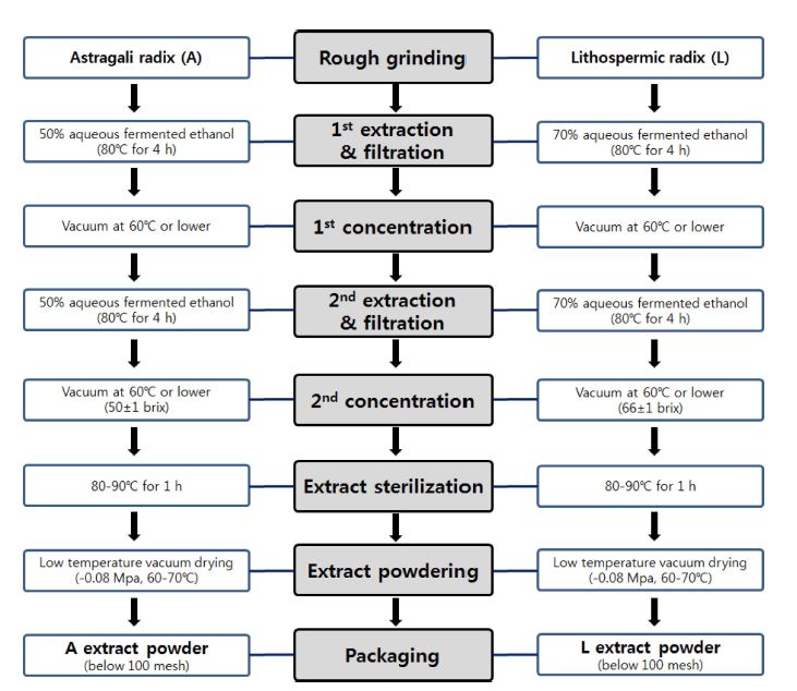
황기 및 지치 건재 시료를 추말로 직경 0.5㎝ 이하의 크기로 조분쇄한 후, 황기 및 지치 분쇄 시료는 유효성분 함량 및 수율 등 선행연구를 바탕으로 각각 50% 주정과 70% 주정을 이용하여 시료 중량의 15 배 용량에서 80℃, 4 시간, 총 2회 환류추출 하였다.
각 추출물은 1㎛ 필터로 여과하여 일차적으로 감압농축하여 최대한 용매를 제거하였다. 이때 황기 추출물, 지치 추출물의 brix는 각각 50 ± 1 brix, 66 ± 1 brix 이었다. 황기 및 지치 추출물은 80 - 90℃에서 1 시간 살균한 후 건조하여 각각 황기추출물 분말 (A extract powder, yield 24%)과 지치 추출물분말 (L extract powder, yield 42%)을 얻었다 (Fig. 1). 기능성 식품으로서의 산업적 활용성을 고려할 때 단가와 원료수급면에서 훨씬 유리한 작물인 황기의 비율이 높은 혼합물을 연구대상으로 하고, A extract powder와 L extract powder의 최적 혼합비를 설정하기 위해 대표적으로 5 : 5, 6 : 4, 7 : 3, 8 : 2의 4 종의 중량비 (w/w)로 혼합한 복합물을 MMPs 저해능 평가 시료로 사용하였다.
3. 세포배양
SW1353 인간 연골세포주는 American Type Culture Collection (ATCC HTB-94, Rockville, MD, USA)으로부터 구입하였으며, 10% FBS와 1% penicillin-streptomycin이 함유된 DMEM 배지에서 37℃, 5% CO2 조건의 배양기에서 48시간 동안 배양하였다. 0.25% (w/v) trypsin 0.53 mM EDTA 용액으로 처리하여 2 일 간격으로 계대 배양하여 실험에 사용하였다.
4. 세포독성 평가
SW1353세포에 대한 각 시료의 세포 독성 (cytotoxicity)을 조사하기 위해 각 농도의 시료를 세포에 처리한 후 MTT assay를 실시하였다. SW1353 (8 × 10⁶ cells/well) 세포를 96 well plate에 분주하여 24 시간 배양하였다. 황기, 지치 추출물 및 혼합 비율별 복합물은 50 - 400㎍/㎖, 단일 성분은 표준품을 이용하여 5 - 80 μM 농도로 각각 세포에 처리한 후, 24 시간 배양하였다. 시료 처리 후 배양액을 제거하고 최종농도가 1㎎/㎖이 되도록 MTT solution을 첨가한 후, 37℃에서 5% CO2 조건으로 2 시간 반응시켰다. 이후, 생성된 formazan crystal을 DMSO에 녹여 microplate reader를 이용하여 570㎚에서 흡광도를 측정하였다. 세포 생존율은 아래의 계산식으로 정상 대조군 (Con) 대비 상대적 생존율을 백분율로 나타내었다.
세포 생존율 (%) = (시료 처리군의 흡광도)/(정상 대조군의 흡광도) × 100
5. Matrix metalloproteinases (MMPs) 활성 측정
연골세포를 6-well 플레이트에 1 × 10⁶ cells/well 농도로 분주하여 24 시간 배양하였다. 세포독성 평가에 사용한 각 시료를 농도별로 30 분간 전처리한 후, IL-1β를 20 ng/㎖로 처리하고 24 시간 동안 반응시켰다. MMP 활성 측정은 형광 ELISA 키트 (SensoLyte® 520 MMPs assay kit, AnaSpec Inc., Fremont, CA, USA)를 이용하여 정량 분석하였다. 형광 ELISA 정량 분석은 세포 배양액에 MMP-1 (71150, AnaSpec Inc., Fremont, CA, USA), MMP-3 (71152, AnaSpec Inc., Fremont, CA, USA)와 MMP-13 (71156, AnaSpec Inc., Fremont, CA, USA) 기질을 처리하여 30 분간 효소반응 후, excitation/emission 490/520㎚에서 측정하였다.
6. 유효성분의 HPLC 분석
HPLC 분석은 YMC의 ODS-AM (4.6 × 250㎜, 5㎛) 컬럼을 사용하여 30℃에서 실시하였다. 이동상 조건은 0.1% formic acid, acetonitrile 용매를 혼합하여 사용하였다. 분석시간은 acetonitrile을 3 분 동안 5%로 유지한 후, 3 분 동안 20%로 증가시키고, 이어서 25 분 동안 28%로 증가시켰다. 이후 4 분 동안 100%로 증가시키는 조건으로 기울기 용리하였다. 유속은 1㎖/min으로 유지하고, UV 검출기의 254㎚ 파장에서 흡광도를 측정하여 황기 유효성분 calycosin과 calycosin-7-O-β-D-glucoside 및 지치 유효성분 lithospermic acid의 함량을 분석하였다.
7. HPLC 분석법 검증 (Validation)
표준정량곡선 작성을 위해 표준물질 calycosin, calycosin-7-O-β-D-glucoside 및 lithospermic acid를 methanol에 녹여 표준용액을 제조하였다. 분석법을 검증하기 위해 직선성(linearity), 검출한계 (limit of detection, LOD) 및 정량한계(limit of quantitaion, LOQ)를 분석하였다. 각 성분에 대한 LOD, LOQ는 표준정량곡선을 작성한 후, 시그널 (S) 대비 노이즈 (N) 값인 S/N 값이 각각 3, 10 일 때의 농도로서 구하였다. 또한 각 성분에 대하여 3 종의 농도 조건을 5 일 동안 반복 분석하여 inter-day precision을 평가하고, 연속으로 5 회 분석하여 intra-day precision를 평가하여 정밀도(precision) 및 정확도 (accuracy)를 측정하고 분석법의 재현성 (reproducibility)을 검증하였다.
8. 통계분석
모든 분석 자료는 평균 ± 표준오차 (means ± SD)로 나타내었으며, 실험결과는 GraphPad Prism® Version 5.0 (GraphPad Software, San Diego, CA, USA) 프로그램을 이용하여 분산분석 (ANOVA)으로 Dunnett’s test 방법을 이용하여 유의성을 검정하였다 (p < 0.05).
결과 및 고찰
황기, 지치 복합물의 matrix metalloproteinases (MMPs) 억제효과
SW1353 세포에 대한 세포독성을 평가하기 위해, SW1353 세포에 황기 (Astragali Radix; Astragalus membranaceus Bunge), 지치 추출물 및 혼합 비율별 복합물을 농도별로 처리한 후, 세포 생존율 (cell viability)을 측정하였다. 모든 시료 처리군에서 아무것도 처리하지 않은 정상군 (Con)과 비교하여 최고 농도 400㎍/㎖ 수준까지 세포 독성을 나타내지 않는 것을 확인하였다 (Fig. 2). 이후 효능검정 시료의 세포 처리농도는 보다 안전한 100과 200㎍/㎖ 수준에서 MMPs 억제 효과를 검정하였다.
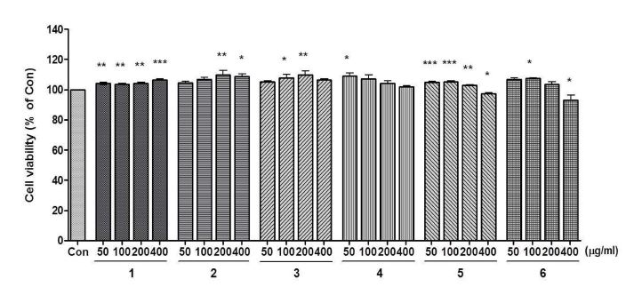
Cell cytotoxicity of extracts and mixtures in different ratio on SW1353 cells.The cells were treated with samples (50 - 400 ㎍/㎖) for 24 h. Cell viability was measured by MTT assay. 1; astragali radix extract (A), 2; lithospermi radix extract (L), 3 - 6; mixtures (A : L ratio, 5 : 5, 6 : 4, 7 : 3 and 8 : 2, respectively). Data were analyzed by One-way ANOVA Dunnett’s test to compare all of the tested groups. *p < 0.05; **p < 0.01; ***p < 0.001 compared with control (100%) cells.
Zhou 등 (2018)의 연구에서는 황기의 열수 추출 후 100% 에탄올 추출물은 3 가지 유방암 세포에 대해서 50㎍/㎖ 수준에서부터 세포 독성을 통해 항암 활성이 있음을 보고하였다. 이를 통해 황기는 세포에 특이적으로 세포 독성을 나타낼 수 있으며, 70% 주정 추출물은 SW1353 연골세포에서는 고농도 수준까지 세포 독성이 없는 것으로 생각된다.
또한, 사전 연구에서 지치의 초임계 추출법을 이용한 추출물은 SW1353 세포에서 5㎍/㎖ 이상의 농도를 처리했을 때 세포 독성이 나타나는 것으로 보고되었다 (Kim et al., 2013a). 하지만 70% 주정을 이용한 환류추출을 했을 때, SW1353 세포에서 고농도 수준까지 세포 독성을 나타내지 않는 것을 확인하였다. 이러한 결과를 통해 지치는 추출법에 의해 세포 독성이 나타날 수 있으며, 70% 주정 추출물은 상대적으로 안전성이 높은 것으로 사료된다. 각각의 황기 또는 지치 추출물과 혼합 비율별 복합물 처리가 연골세포주에서 IL-1β에 의해 유도된 MMP-1, MMP-3 및 MMP-13의 생성량 증가에 대한 억제 효과를 나타내는지 평가하였다. IL-1β 처리에 따른 MMP-1의 생성량 변화를 확인한 결과, IL-1β 처리군은 정상대조군 (Con)과 비교하여 MMP-1의 생성량이 약 9.6 배 증가하였다 (Fig. 3).
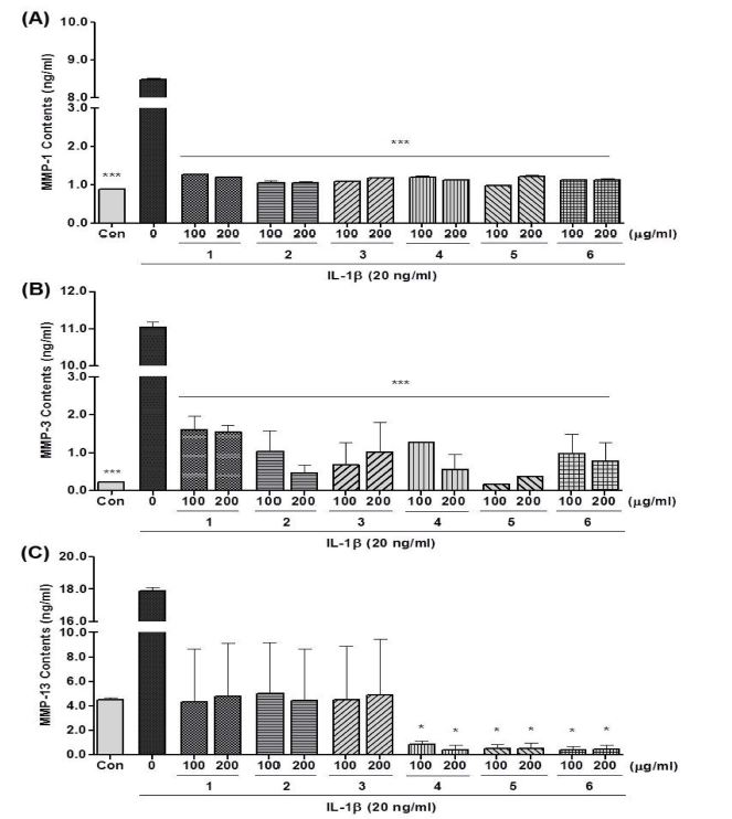
Inhibitory effects of mixtures on the levels of (A) MMP-1, (B) MMP-3, and (C) MMP-13 in IL-1β-treated SW1353 cells.1; astragali radix extract (A), 2; lithospermi radix extract (L), 3 - 6; mixtures (A : L ratio, 5 : 5, 6 : 4, 7 : 3 and 8 : 2, respectively). Data were analyzed by One-way ANOVA Dunnett’s test to compare all of the tested groups. *p < 0.05; **p < 0.01; ***p < 0.001 compared with only IL-1β treated cells.
그러나 시료를 처리했을 경우, 모든 시료 처리군에서 증가된 MMP-1의 생성량이 정상 대조군과 유사한 수준까지 감소하는 것을 확인하였다. 이러한 결과는 황기 및 지치 단일 추출물이 MMP-1 생성량 증가에 대한 강력한 억제 효과가 있는 것으로 생각되며, 다양한 혼합 비율에 의한 복합물의 경우에도 단일 추출물의 억제 효과가 감소되지 않고 발휘되었기 때문에 복합물 간의 활성 정도가 유사하게 나타난 것으로 사료된다.
MMP-3의 생성량 변화의 경우, IL-1β 처리군은 정상 대조군(Con)과 비교하여 MMP-3의 생성량이 약 49.9 배 증가하였다. 그러나 시료를 처리했을 경우, 모든 시료 처리군에서 IL-1β 처리에 의한 MMP-3의 생성량이 감소되는 것을 확인하였다. 특히, 7 : 3 혼합 비율의 복합물은 100㎍/㎖ 처리시, IL-1β 처리군 대비하여 98.5% 감소 효과를 보였으며, 모든 시료 중 가장 효과적으로 MMP-3의 생성 증가를 억제하였으며, 동일 농도의 황기 추출물 (85.4%) 및 지치 추출물 (90.7%) 처리군과 비교하여 더 효과적으로 MMP-3 생성을 억제하는 것을 확인하였다. 이러한 결과는 두 가지 단일 추출물이 혼합되어 시너지 작용에 의해 MMP-3 생성 억제 효과가 증대된 것으로 사료된다.
MMP-13의 생성량 변화에서는 IL-1β 처리군은 정상 대조군(Con)과 비교하여 MMP-13의 생성량이 약 4.0 배 증가한 것을 확인하였다. 그러나 시료를 처리했을 경우, 모든 시료 처리군에서 증가된 MMP-13의 생성량이 감소된 것을 확인하였다. 시료들 중에서 6 : 4, 7 : 3 및 8 : 2 혼합 비율의 복합물은 100㎍/㎖ 처리시, IL-1β 처리군과 비교하여 각각 95.2, 97.1 및 97.6% MMP-13 생성량 감소 효과를 보였다. 동일 농도의 단일 추출물 처리군에서는 IL-1β 처리군과 비교하여 각각 75.7, 72.0% 억제 효과를 보였으며, 이러한 결과를 통해 단일 추출물 처리군에 비해 6 : 4, 7 : 3 및 8 : 2 혼합물에서 시너지 효과를 나타낼 수 있음을 확인하였다.
이러한 결과를 통해 황기, 지치 단일 추출물 및 복합물이 IL-1β 처리에 의한 연골세포의 MMP-1, MMP-3 및 MMP-13의 생성량 증가를 억제하는 효과가 있는 것을 확인하였다. 하지만, 복합물의 경우 혼합 비율 및 MMPs 종류에 따라 MMPs 생성 억제 효과는 단일 추출물과 비교하여 증폭 또는 감소하여 서로 상이한 결과를 나타내었으며, 혼합 비율-의존적인 효과는 관찰되지 않았다. 특히, 7 : 3 혼합 비율의 복합물은 황기 또는 지치 단일 추출물 및 다른 혼합 비율의 복합물과 비교하여 IL-1β 처리에 의한 연골세포의 MMP-1, MMP-3 및 MMP-13의 생성량 증가를 가장 효과적으로 억제하였으며, 이는 각각의 단일 추출물이 가지고 있는 MMPs 생성 억제 효능이 복합되어 시너지 작용 (synergistic actions)에 의해 증폭효과를 보인 것으로 생각된다. 따라서 추후 황기, 지치 복합물을 이용한 골관절염 개선 연구에는 7 : 3 (황기 :지치, w/w) 비율이 최적의 혼합 조성비로 사료된다.
MMPs는 관절염 발병 과정에서 염증 반응에 의해 연골 기질이 분해되는데 관여하며, 그 중에서도 MMP-1, MMP-3 및 MMP-13은 골관절염 환자에서 높게 발현되어 있으며 MMPs의 저해는 골관절염 치료에서 핵심적 역할을 한다고 보고되었다 (Jin et al., 2001; Blaney Davidson et al., 2009; Li et al., 2011). Kwon 등 (2015)의 연구에 의하면 참당귀 추출물이 연골세포와 골관절염 동물 모델에서 MMPs의 활성을 효과적으로 억제하여 골관절염의 발생을 억제하는 효과가 있는 것으로 보고하였다. 또한, Choi 등 (2017)의 연구에서는 아세틸콜린에스터라제 활성과 지질 산화에 대해서 계피 및 금앵자를 혼합한 복합물이 단일 추출물보다 더 효과적으로 억제하였으며, 이는 복합물이 시너지 효과를 나타내는 것으로 보고하였다.
따라서 황기 및 지치 추출물을 7 : 3 비율로 혼합한 복합물은 시너지 효과를 통해 MMP-1, MMP-3 및 MMP-13의 활성 또는 생성 증가를 저해하여 연골세포 보호 효과 및 골관절염의 발생에 대한 억제 효과를 나타낼 수 있을 것으로 예상된다.
2. 유효성분의 MMPs 억제효과
황기 복합물의 MMPs 생성 억제 효과에 대한 유효성분을 확인하고자 황기 또는 지치 유래 단일 성분을 선정하여 세포독성 및 MMPs 생성 억제능을 평가하였다. 황기는 주요 flavonoid 성분인 calycosin 및 calycosin-7-O-β-D-glucoside를 선정하여 활성을 평가하였다. 최근 황기의 total flavonoid 성분의 in vivo 및 in vitro에서의 면역조절 및 항염증 효과가 보고된 바 있다 (Guo et al., 2016). 또한 calycosin-7-O-β-D-glucoside는 선행연구에서 골관절염에 대한 효과가 보고되었지만 (Choi et al., 2007), 연골세포 보호 효과 및 그 기전에 대한 연구는 보고되지 않았다.
지치는 shikonin 계열의 색소 성분의 항염증 활성이 보고되었으며, 우리는 선행연구에서, 지치로부터 분리한 pseudoshikonin 성분이 연골세포주에서 MMPs 효소 저해능을 나타냄을 보고한 바 있다 (Lee et al., 2016). 하지만 shikonin 계 색소성분은 비극성 naphthoquinone 계열의 성분인 반면 본 실험의 지치 추출물은 70% 주정 추출물이기 때문에 lithospermic acid를 유효성분으로 선정하여 활성을 평가하였다. Lithospermic acid는 지치에서 발견되는 주요 phenylpropanoid oligomer 성분이며 (Murata et al., 2013), 항산화, 간 보호 효과 및 심혈관계 질환에서 항섬유화 효과 등 다양한 생리활성이 있다고 알려져 있다 (Shigematsu et al., 1994; Chan and Ho, 2015).
황기 및 지치 유래 3 가지 단일 성분의 SW1353 세포에 대한 독성을 평가한 결과, 3 가지 단일 성분 모두 최대 농도 80 μM 수준까지 정상 대조군 (Con)과 비교하여 독성을 나타내지 않는 것을 확인하였다 (Fig. 4).
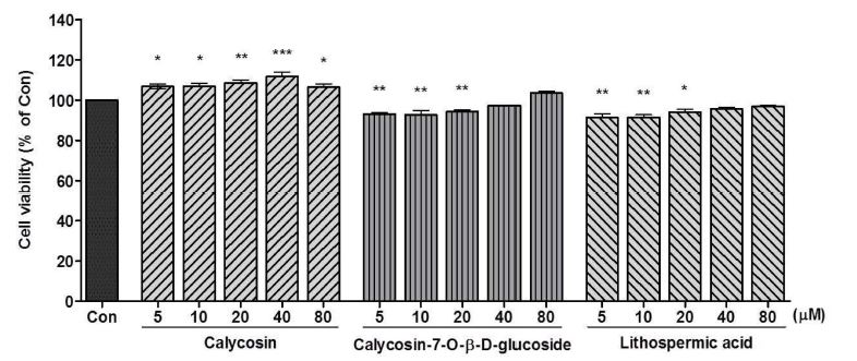
Cytotoxicity of calycosin, calycosin-7-O-β-D-glucoside and lithospermic acid from Astragali radix or Lithospermi radix in SW1353 cells.The cells were treated with three compounds (5 - 80 μM) for 24 h. Cell viability was determined by MTT assay. Data were analyzed by One-way ANOVA Dunnett’s test to compare all of the tested groups. *p < 0.05; **p < 0.01; ***p < 0.001 compared with control (100%) cells.
3 가지 단일 성분이 연골세포주에서 IL-1β에 의해 유도된 MMP-1, MMP-3 및 MMP-13의 생성량 증가에 미치는 영향을 평가한 결과, calycosin 40 μM 처리군은 IL-1β 처리군에 대비하여 MMP-1 및 MMP-3 생성량을 각각 19.0 및 43.7% 감소시켰다. 그러나, MMP-13의 생성 억제 효과는 거의 나타나지 않았다 (Fig. 5A). Calycosin-7-O-β-D-glucoside 40 μM 처리시, IL-1β 처리군과 비교하여 MMP-1, MMP-3 및 MMP-13 생성량이 각각 20.3, 54.6 및 34.5% 감소하는 것을 확인하였다 (Fig. 5B).
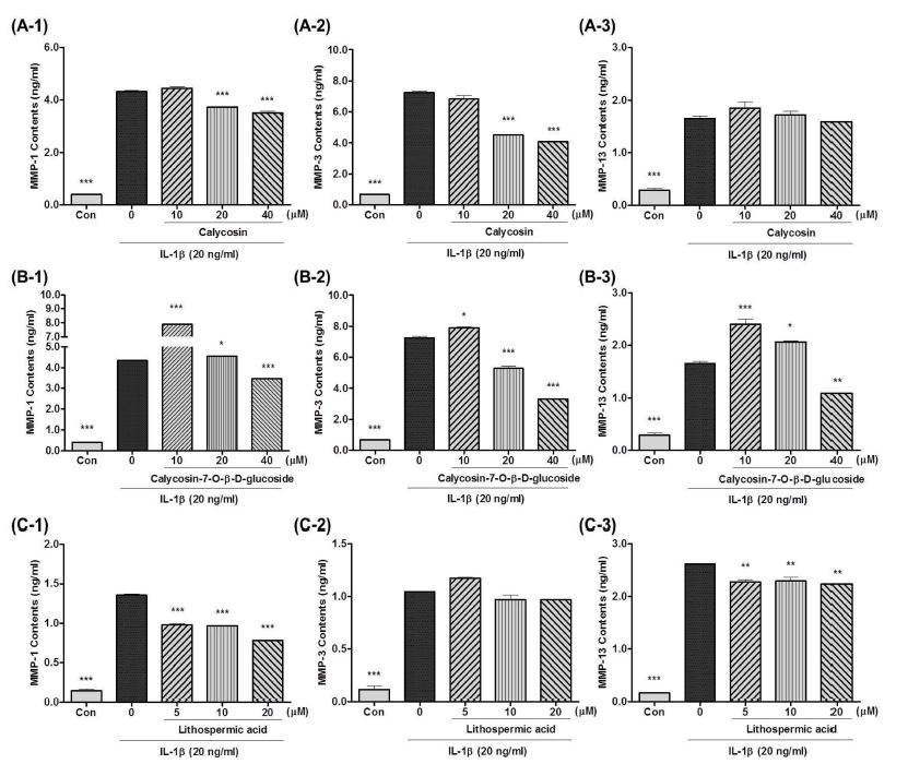
Effects of (A) calycosin, (B) calycosin-7-O-β-D-glucoside and (C) lithospermic acid on MMP-1, MMP-3 and MMP-13 production in IL-1β-treated SW1353 cells.(A-1, B-1 and C-1); MMP-1, (A-2, B-2 and C-2); MMP-3, (A-3, B-3 and C-3); MMP-13 contents. *Data were analyzed by One-way ANOVA Dunnett’s test to compare all of the tested groups. *p < 0.05; **p < 0.01; ***p < 0.001 compared with only IL-1β treated cells.
최근, 황기의 total flavonoid 성분이 Freund's complete adjuvant (FCA)로 유도된 동물 모델에서 관절의 활액 조직의 osteoprotegerin (OPG), receptor activator of nuclear factor-κB ligand (RANKL) 및 nuclear factor (NF)-κB p65 발현의 조절에 의한 항관절염 효과가 있는 것으로 보고되었다 (Liu et al., 2017). OPG/RANKL/RANK은 중요한 골대사의 신호 조절 시스템이자 파골세포형성 (osteoclastogenesis)에서 중요한 기능을 하는 것으로도 알려져 있어 (Wada et al., 2006), 황기의 flavonoid 성분은 골관절염, 류마티스 관절염 등 모든 관절염의 개선 및 관절건강에 활용성이 높은 기능성 성분임을 알 수 있다.
또한, 지치 유래 lithospermic acid 20 μM 처리시, IL-1β 처리군 대비하여 MMP-1의 생성량 증가에 대하여 42.7%의 저해 효과를 보였으며, MMP-3 및 MMP-13의 생성량에 대해서는 감소 효과가 각각 7.0 및 14.4%로 거의 없는 것으로 확인되었다 (Fig. 5C).
지치 주정추출물에 함유되어 있는 lithospermic acid는 통풍성 관절염 모델에서 항염증 효과 및 혈당저하 효과를 갖는 것으로 보고된 바 있으나 (Liu et al., 2008), 관절의 연골보호 효과 및 그 작용기전에 대한 연구는 아직 보고되지 않았다. 다만, 지치 메탄올 추출물이 피부 콜라겐의 분해를 저해함으로써 산화적 스트레스로 유도된 피부노화를 억제하고 특히, 인간의 피부 섬유아세포에서 MMP-1의 발현을 감소시키며, 이는 지치의 phenol 화합물의 작용에 기인할 수 있다고 언급한 바 있다 (Yoo et al., 2014).
이러한 결과를 통해, calycosin-7-O-β-D-glucoside, calycosin 및 lithospermic acid는 MMP-1, MMP-3 및 MMP-13 생성을 효과적으로 억제하는 것으로 확인되었다. 이로써 이들 성분이 황기 복합물의 연골보호 효능과 품질을 평가하는 주요 기능성분이면서 지표성분으로 활용할 수 있을 것으로 사료된다.
3. 유효성분의 HPLC 분석
황기, 지치 복합물의 유효성분으로 선정하여 MMPs 저해능을 평가한 calycosin, calycosin-7-O-β-D-glucoside와 lithospermic acid에 대하여 황기 복합물에서의 함량을 정량하기 위해, 이들 3 종의 성분을 동시 분석할 수 있는 HPLC 분석법을 검토하였다.
선행연구에서 황기 단독 추출물의 기능성 원료 개발을 위한 지표성분 분석법 제시를 위해 황기 지표성분에 대한 분석법 검증을 실시한 바 있으나 (Kim et al., 2013b), 이번 연구는 황기, 지치 복합물의 기능성분을 동시에 분석하는 다성분 동시 분석법으로서 보다 정밀하고 진보된 분석법 검토가 필요하였다. 최종적으로, YMC의 ODS-AM 역상 컬럼을 사용하고, H2O와 acetonitrile의 혼합용매로 기울기 용리하여 254㎚ 파장에서 흡광도를 측정한 결과, calycosin, calycosin-7-O-β-D-glucoside, lithospermic acid 각각의 성분이 양호한 분리능을 나타내는 HPLC 동시 분석법을 확립하였다 (Fig. 6). 이러한 HPLC 분석법을 검증하기 위해, 0.78 - 100 ng/㎕ 농도 범위 내에서 분석하여 얻은 피크면적을 이용하여 회귀식과 상관계수를 각각 구하였다.
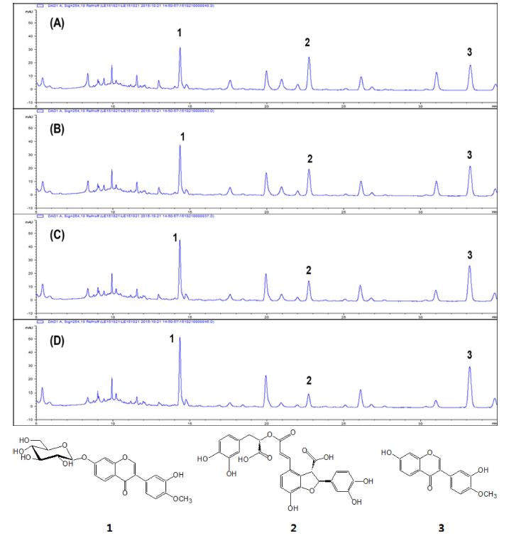
HPLC chromatogram of the mixtures with a composition ratio [Astragali radix extract (A) : Lithospermi radix extract (L), w/w].(A); 5 : 5, (B); 6 : 4, (C); 7 : 3 and (D); 8 : 2. The structures of calycosin-7-O-β-D-glucoside (1), lithospermic acid (2) and calycosin (3).
Table 1에서와 같이 calycosin, calycosin-7-O-β-D-glucoside과 lithospermic acid의 회귀식을 나타내었으며, 이들 성분의 상관계수 (R2) 값은 모두 0.999 로서 높은 직선성을 나타내었다. Signal 대비 noise 즉, S/N 비에 근거한 calycosin, calycosin-7-O-β-D-glucoside 및 lithospermic acid의 최저 검출한계농도 (LOD)는 각각 1.06, 0.56, 2.45 ng/㎕이었고, 최저 정량한계농도 (LOQ)는 4.50, 2.88, 8.22 ng/㎕로 측정되었다.
HPLC 분석법의 재현성 검정과 유효성분의 회수율 분석은 다음과 같이 실시하였다. 각각의 유효성분에 대해 10, 25, 50 ng/㎕의 3 가지 농도에서 하루 동안의 분석 재현성 (5 회 반복, intra-day)과 연속 5 일 동안의 분석 재현성 (5 회 반복, inter-day)을 실시하고, 정밀도 (precision) 및 정확도 (accuracy)는 각각 상대표준오차 값과 회수율로서 나타내었다 (Table 2).
유효성분 3 종의 intra-day 정밀도는 0.52 - 1.69%, 정확도는 98.48 - 101.36% 범위였으며, inter-day 정밀도와 정확도는 각각 0.33 - 2.02%, 96.51 - 100.24% 범위로 측정되었다. Intra-day와 inter-day 분석 결과는 주어진 농도 범위 내에서 약 15%를 벗어나지 않는 정확성을 보여주었다.
Calycosin-7-O-β-D-glucoside와 lithospermic acid는 약리학적 이용을 위해 각각 rat의 plasma와 serum에서 분석법이 검증된 바 있는데, HPLC-MS를 이용한 분석법 보고에서 calycosin-7-O-β-D-glucoside은 10, 100, 1,000 ng/㎖의 농도 수준에서 정밀도 측정시, inter-day 및 intra-day 결과가 각각 1.9 - 4.46%, 1.66 - 3.14%로 분석되어 본 연구의 분석법보다는 정밀도가 다소 떨어지는 경향을 보였다 (Wen et al., 2008).
또한 lithoseprmic acid는 LC/MS/MS를 이용한 분석법 검증에서 inter-day에서 7.94 - 9.37%의 정밀성을 나타내었다(Wang et al., 2008). 결론적으로, 이번 HPLC 분석법이 비교적 높은 정확도와 정밀도를 보여줌에 따라 황기, 지치 복합물의 유효성분 HPLC 분석법으로 적합하다고 판단되었다.
이 HPLC 분석법을 사용하여 황기, 지치 추출물 및 이들 추출물을 5 : 5, 6 : 4, 7 : 3, 8 : 2의 혼합비로 제조한 복합물에서 유효성분 3 종의 함량을 정량 분석하였다. Table 3에서와 같이 황기 단독 추출물의 calycosin과 calycosin-7-O-β-D-glucoside 함량은 각각 0.603, 0.818㎎/g 수준이었으며, 지치 단독 추출물의 lithospermic acid 함량은 1.78㎎/g 수준이었다. 황기, 지치 복합물은 혼합 비율에 따라 calycosin 함량은 0.304 - 0.483㎎/g, calycosin-7-O-β-D-glucoside 함량은 0.429-0.666㎎/g, lithospermic acid 함량은 0.382 - 0.912㎎/g 수준이었으며, 가장 우수한 MMPs 저해 활성을 나타낸 7 : 3 혼합비의 복합물은 calycosin, calycosin-7-O-β-D-glucoside 및 lithospermic acid 함량이 각각 0.424, 0.586, 0.554㎎/g으로 분석되었다.

Quantification of index compounds in individual extracts and mixtures of the different mixing ratio [Astragali radix extract (A) : Lithospermi radix extract (L)].
본 연구 결과를 종합해 볼 때, 황기, 지치의 주정 추출물 제조공정을 확립하였으며, 연골세포에서의 MMP-1, MMP-3 및 MMP-13 생성 억제효과 평가를 통해 황기와 지치 추출물의 최적 복합 조성비를 결정하고, 혼합에 의한 시너지 효과(synergistic effect)를 확인하였다.
추가적으로 황기 유래 calycosin-7-O-β-D-glucoside, calycosin과 지치 유래 lithospermic acid는 MMP-1, MMP-3 및 MMP-13의 생성을 억제하는 효과를 나타냈으며, 황기, 지치 복합물의 연골보호 효과에 대한 주요 유효성분으로 작용한다고 사료된다. 혼합 비율 7 : 3의 황기, 지치 복합물은 우수한 MMPs 생성 억제 활성을 나타내어 이 복합물을 활용한 골관절염 동물 모델을 이용한 효능평가 연구 및 분자생물학적인 방법을 통한 기전 연구 등의 기초자료로 활용될 것으로 판단된다. 이러한 심도 깊은 연구를 통해 황기, 지치 복합물은 관절건강에 대한 기능성 원료로 개발될 가능성이 높다고 사료된다.
Acknowledgments
본 연구는 농촌진흥청 연구사업(과제번호: PJ01329101)의 지원에 의해 이루어진 결과로 이에 감사드립니다.
References
-
Aigner, T, and McKenna, L, (2002), Molecular pathology and pathobiology of osteoarthritic cartilage, Cellular and Molecular Life Sciences, 59, p5-18.
[https://doi.org/10.1007/s00018-002-8400-3]

-
Blaney Davidson, EN, Remst, DFG, Vitters, EL, van Beuningen, HM, Blom, AB, Goumans, MJ, van den Berg, WB, and van der Kraan, PM, (2009), Increase in ALK1/ALK5 ratio as a cause for elevated MMP-13 expression in osteoarthritis in humans and mice, Journal of Immunology, 182, p7937-7945.
[https://doi.org/10.4049/jimmunol.0803991]

-
Chan, KWK, and Ho, WS, (2015), Anti-oxidative and hepatoprotective effects of lithospermic acid against carbon tetrachloride-induced liver oxidative damage in vitro and in vivo, Oncology Reports, 34, p673-680.
[https://doi.org/10.3892/or.2015.4068]

-
Chen, JJ, Huang, JF, Du, WX, and Tong, PJ, (2014), Expression and significance of MMP3 in synovium of knee joint at different stage in osteoarthritis patients, Asian Pacific Journal of Tropical Medicine, 7, p297-300.
[https://doi.org/10.1016/s1995-7645(14)60042-0]

-
Cho, WCS, and Leung, KN, (2007), in vitro and in vivo anti-tumor effects of Astragalus membranaceus, Cancer Letters, 252, p43-54.
[https://doi.org/10.1016/j.canlet.2006.12.001]

- Choi, SI, Heo, TR, Min, BH, Cui, JH, Choi, BH, and Park, SR, (2007), Alleviation of osteoarthritis by calycosin-7-O-β-D-glucopyranoside(CG) isolated from Astragali radix(AR) in rabbit osteoarthritis(OA) model, Osteoarthritis and Cartilage, 15, p1086-1092.
- Choi, SJ, Kim, CR, Park, CK, Gim, MC, Choi, JH, and Shin, DH, (2017), Ameliorating effects of Cinnamomum loureiroi and Rosa laevigata extracts mixture against trimethyltin-induced learning and memory impairment model, Korean Journal of Medicinal Crop Science, 25, p353-360.
-
Goldring, MB, (2000), The role of the chondrocyte in osteoarthritis, Arthritis and Rheumatology, 43, p1916-1926.
[https://doi.org/10.1002/1529-0131(200009)43:9<1916::aid-anr2>3.0.co;2-i]

-
Guo, Z, Xu, HY, Xu, L, Wang, SS, and Zhang, XM, (2016), in vivo and in vitro immunomodulatory and anti-inflammatory effects of total flavonoids of astragalus, African Journal of Traditional, Complementary and Alternative Medicines, 13, p60-73.
[https://doi.org/10.21010/ajtcam.v13i4.10]

- Hong, EY, Kyung, MJ, and Whang, WK, (2015), Isolation of constituents of Lithospermum erythrorhizon and development of an analytical method of its constituents by HPLC-UV, Yakhak Hoeji, 59, p222-229.
- Hwang, SY, Hwang, BY, Kang, SS, Kim, CM, Park, JH, Bae, KH, Sun, KH, Lee, SH, Chang, SY, Kang, SJ, Ro, JS, and Lee, KS, (2000), Isolation and quantitative analysis of acetylshikonin from Lithospermi radix, Korean Journal of Pharmacognosy, 31, p295-299.
- Im, KR, Kim, MJ, Jung, TK, and Yoon, KS, (2010), Analysis of isoflavonoid contents in Astragalus membranaceus Bunge cultivated in different areas and at various ages, Korean Society for Biotechnology and Bioengineering Journal, 25, p271-276.
- Jin, SY, Kang, SS, Lee, DW, Yim, SJ, Yun, YH, and Lee, BI, (2001), Expression of matrix metalloproteinase(MMP)-1, MMP-3 nitrotyrosine and apoptosis in articular cartilage of human osteoarthritis, Korean Journal of Pathology, 35, p319-329.
-
Kim, G, Choi, S, Lee, D, Noh, H, Lee, S, Choi, J, and Kim, S, (2014), Effects of ethanol extracts and compounds from Astragali radix on chondrocytes and MIA-induced osteoarthritis model in rat, Planta Medica, 80(P2B35), https://www.thieme-connect.com/products/10.1055/s-0034-1394912 (cited by 2019 June 5).
[https://doi.org/10.1055/s-0034-1394912]

- Kim, GS, Kim, HJ, Lee, DY, Choi, SM, Lee, SE, Noh, HJ, Choi, JG, and Choi, SI, (2013a), Effects of supercritical fluid extract, shikonin and acetylshikonin from Lithospermum erythrorhizon on chondrocytes and MIA-induced osteoarthritis in rats, Korean Journal of Medicinal Crop Science, 21, p466-473.
- Kim, GS, Lee, DY, Lee, SE, Noh, HJ, Choi, JH, Park, CG, Choi, SI, Hong, SJ, and Kim, SY, (2013b), Evaluation on extraction conditions and HPLC analysis method for bioactive compounds of Astragali radix, Korean Journal of Medicinal Crop Science, 21, p486-492.
-
Kim, GS, Park, CG, Lee, KH, Choi, JH, Lee, SE, Noh, HJ, Lee, JH, and Kim, SY, (2011), Investigation of shikonin pigments and antioxidant activity of the roots from Lithospermum erythrorhizon according to the different growth stages and areas of cultivation, Korean Journal of Medicinal Crop Science, 19, p435-440.
[https://doi.org/10.7783/kjmcs.2011.19.6.435]

- Kim, JS, Han, YS, and Kang, MH, (2006), Identification of shikonin and its derivatives from Lithospermum erythrorhizon, Journal of the Korean Society of Food Science and Nutrition, 35, p177-181.
-
Koo, HJ, Sohn, EH, and Kang, SC, (2013), The optimal combination of the mixture of unripe Rubus coreanus and Astragalus membranaceus in the activation and differentiation of osteoblastic cells, Korean Journal of Plant Resources, 26, p658-662.
[https://doi.org/10.7732/kjpr.2013.26.5.658]

-
Kwon, JH, Han, MS, and Lee, YM, (2015), Regulatory mechanism of Angelica Gigas extract powder on matrix metalloproteinases in vitro and in vivo model, Analytical Science and Technology, 28, p361-369.
[https://doi.org/10.5806/ast.2015.28.6.361]

-
Lee, DY, Choi, SI, Han, SH, Lee, YJ, Choi, JG, Lee, YS, Choi, JH, Lee, SE, and Kim, GS, (2016), Potential of pseudoshikonin I isolated from Lithospermi radix as inhibitors of MMPs in IL-1β-induced SW1353 cells, International Journal of Molecular Sciences, 17, p1350, (cited by 2019 May 14).
[https://doi.org/10.3390/ijms17081350]

- Lee, HH, Bai, S, and Chin, JE, (2005), Inhibitory effect of Lithospermum erythrorhizon extracts on melanin biosynthesis, Journal of the Korean Society of Food Science and Nutrition, 34, p1325-1329.
-
Li, NG, Shi, ZH, Tang, YP, Wang, ZJ, Song, SL, Qian, LH, Qian, DW, and Duan, JA, (2011), New hope for the treatment of osteoarthritis through selective inhibition of MMP-13, Current Medicinal Chemistry, 18, p977-1001.
[https://doi.org/10.2174/092986711794940905]

-
Liu, X, Chen, R, Shang, Y, Jiao, B, and Huang, C, (2008), Lithospermic acid as a novel xanthine oxidase inhibitor has anti-inflammatory and hypouricemic effects in rats, Chemico-Biological Interactions, 176, p137-142.
[https://doi.org/10.1016/j.cbi.2008.07.003]

-
Liu, XY, Xu, L, Wang, Y, Li, JX, Zhang, Y, Zhang, C, Wang, SS, and Zhang, XM, (2017), Protective effects of total flavonoids of Astragalus against adjuvant-induced arthritis in rats by regulating OPG/RANKL/NF-κB pathway, International Immunopharmacology, 44, p105-114.
[https://doi.org/10.1016/j.intimp.2017.01.010]

- Mandelbaum, B, and Waddell, D, (2005), Etiology and pathophysiology of osteoarthritis, Orthopedics, 28, p207-214.
- Murata, T, Oyama, K, Fujiyama, M, Oobayashi, B, Umehara, K, Miyase, T, and Yoshizaki, F, (2013), Diastereomers of lithospermic acid and lithospermic acid B from Monarda fistulosa and Lithospermum erythrorhizon, Fitoterapia, 91, p51-59.
- Nagase, H, and Woessner, JF, (1999), Matrix metalloproteinases, Journal of Biological Chemistry, 274, p21491-21494.
-
Qin, Q, Niu, J, Wang, Z, Xu, W, Qiao, Z, and Gu, Y, (2012), Astragalus membranaceus extract activates immune response in macrophages via heparanase, Molecules, 17, p7232-7240.
[https://doi.org/10.3390/molecules17067232]

-
Shigematsu, T, Tajima, S, Nishikawa, T, Murad, S, Pinnell, SR, and Nishioka, I, (1994), Inhibition of collagen hydroxylation by lithospermic acid magnesium salt, a novel compound isolated from Salviae miltiorrhizae radix, Biochimica et Biophysica Acta, 1200, p79-83.
[https://doi.org/10.1016/0304-4165(94)90030-2]

-
Shirataki, Y, Takao, M, Yoshida, S, and Toda, S, (1997), Antioxidative components isolated from the roots of Astragalus membranaceus Bunge(Astragali radix), Phytotherapy Research, 11, p603-605.
[https://doi.org/10.1002/(sici)1099-1573(199712)11:8<603::aid-ptr161>3.3.co;2-l]

-
Su, X, Huang, Q, Chen, J, Wang, M, Pan, H, Wang, R, Zhou, Z, Liu, J, Yang, F, Li, T, and Liu, L, (2016), Calycosin suppresses expression of pro-inflammatory cytokines via the activation of p62/Nrf2-linked heme oxygenase 1 in rheumatoid arthritis synovial fibroblasts, Phytotherapy Research, 113, p695-704.
[https://doi.org/10.1016/j.phrs.2016.09.031]

- Tanaka, S, Hamanishi, C, Kikuchi, H, and Fukuda, K, (1998), Factors related to degradation of articular cartilage in osteoarthritis: A review, Seminars in Arthritis and Rheumatism, 27, p392-399.
-
Thuong, PT, Kang, KW, Kim, JK, Seo, DB, Lee, SJ, Kim, SH, and Oh, WK, (2009), Lithospermic acid derivatives from Lithospermum erythrorhizon increased expression of serine palmitoyltransferase in human HaCaT cells, Bioorganic and Medicinal Chemistry Letters, 19, p1815-1817.
[https://doi.org/10.1016/j.bmcl.2009.01.052]

- Toda, S, and Shirataki, Y, (1998), Inhibitory effects of isoflavonoids in roots of Astragalus membranaceus Bunge(Astragali radix) on lipid peroxidation by reactive oxygen species, Phytotherapy Research, 12, p59-61.
-
Wada, T, Nakashima, T, Hiroshi, N, and Penninger, JM, (2006), RANKL-RANK signaling in osteoclastogenesis and bone disease, Trends in Molecular Medicine, 12, p17-25.
[https://doi.org/10.1016/j.molmed.2005.11.007]

-
Wang, L, Zhang, Q, Li, X, Lu, Y, Xue, Z, Xuan, L, and Wang, Y, (2008), Pharmacokinetics and metabolism of lithospermic acid by LC/MS/MS in rats, International Journal of Pharmaceutics, 350, p240-246.
[https://doi.org/10.1016/j.ijpharm.2007.09.001]

- Wang, XJ, and Feng, P, (2000), Antioxidant activity of qizhu tang, Acta Pharmacologica Sinica, 21, p1141-1144.
- Wen, XD, Qi, LW, Li, P, Bao, KD, Yan, XW, Yi, L, and Li, CY, (2008), Simultaneous determination of calycosin-7-O-β-D-glucoside, ononin, astragaloside Ⅳ, astragaloside Ⅰ and ferulic acid in rat plasma after oral administration of Danggui Buxue Tang extract for their pharmacokinetic studies by liquid chromatography-mass spectrometry, Journal of Chromatography B, 865, p99-105.
-
Wu, YS, Hu, YY, Yang, RF, Wang, Z, and Wei, YY, (2007), The matrix metalloproteinases as pharmacological target in osteoarthritis: Statins may be of therapeutic benefit, Medical Hypotheses, 69, p557-559.
[https://doi.org/10.1016/j.mehy.2007.01.042]

-
Yang, ZG, Sun, HX, and Fang, WH, (2005), Haemolytic activities and adjuvant effect of Astragalus membranaceus saponins (AMS) on the immune responses to ovalbumin in mice, Vaccine, 23, p5196-5203.
[https://doi.org/10.1016/j.vaccine.2005.06.016]

-
Yoo, HG, Lee, BH, Kim, W, Lee, JS, Kim, GH, Chun, OK, Koo, SI, and Kim, DO, (2014), Lithospermum erythrorhizon extract protects keratinocytes and fibroblasts against oxidative stress, Journal of Medicinal Food, 17, p1189-1196.
[https://doi.org/10.1089/jmf.2013.3088]

- Yu, DH, Bao, YM, Wei, CL, and An, LJ, (2005), Studies of chemical constituents and their antioxidant activities from Astragalus mongholicus Bunge, Biomedical and Environmental Sciences, 18, p297-301.
-
Zhang, YZ, Xu, F, Dong, J, Liang, J, Hashi, Y, Shang, MY, Yang, DH, Wang, X, and Cai, SQ, (2012), Profiling and identification of the metabolites of calycosin in rat hepatic 9000×g supernatant incubation system and the metabolites of calycosin-7-O-β-D-glucoside in rat urine by HPLC-DAD-ESI-IT-TOF-MSn technique, Journal of Pharmaceutical and Biomedical Analysis, 70, p425-439.
[https://doi.org/10.1016/j.jpba.2012.06.006]

-
Zhou, R, Chen, H, Chen, J, Chen, X, Wen, Y, and Xu, L, (2018), Extract from Astragalus membranaceus inhibit breast cancer cells proliferation via PI3K/AKT/mTOR signaling pathway, BMC Complementary and Alternative Medicine, 18, p83, (cited by 2019 June 10).
[https://doi.org/10.1186/s12906-018-2148-2]

