Anti-inflammatory, Anti-aging, Antioxidant, and Anti-melanogenesis Effects of Extracts from Toona sinensis Leaf
This is an open access article distributed under the terms of the Creative Commons Attribution Non-Commercial License (http://creativecommons.org/licenses/by-nc/3.0/) which permits unrestricted non-commercial use, distribution, and reproduction in any medium, provided the original work is properly cited.
Abstract
Many researchers have developed cosmetic and medicinal ingredients using natural plant extracts to regulate and improve the skin health. We investigated Toona sinensis leaf (TSL) extract as a potential functional cosmetic ingredient by testing its pharmacological effects on skin cells.
The anti-inflammatory and anti-aging effects of TSL were evaluated in tumor necrosis factor (TNF)-α-stimulated human epidermal keratinocytes (HaCaT) and human dermal fibroblasts (HS68) by quantitative reverse transcription polymerase chain reaction and enzyme-linked immunosorbent assay. Notably, TSL antioxidant effects were assessed using 2,2-diphenyl-1-picrylhydrazyl (DPPH) radical scavenging assays. Its anti-melanogenic activity of TSL was determined by quantifying extracellular and intracellular melanin content in α-melanocyte stimulating hormone (α-MSH)-stimulated B16F10 melanoma cells. Furthemore, its inhibitory effects on mushroom tyrosinase activity was investigated. Western blotting was performed to identify the anti-melanogenesis mechanism. Notably, TSL decreased interleukin (IL)-6, IL-8, and matrix metalloproteinase (MMP)-1 expression in TNF-α-stimulated HaCaT and HS68 cells. Furthermore, TSL inhibited DPPH radical activity in a concentration-dependent manner, significantly reduced α-MSH-induced intra/extracellular melanin production in B16F10 cells, and inhibited mushroom tyrosinase activity. Additionally, TSL treatment decreased protein levels of melanogenic enzymes.
Notably, TSL demonstrated anti-inflammatory, anti-aging, antioxidant, and anti-melanogenetic effects on skin cells, confirming its protective effects on skin physiology and suggesting its potential as a functional cosmetic ingredient.
Keywords:
Toona sinensis, Functional Cosmetic Ingredient, Human Dermal Fibroblasts, Human Epidermal Keratinocytes, Inflammation, Matrix Metalloproteinase-1, Melanogenesis, Oxidative StressINTRODUCTION
Skin, the outermost layer of the body consisting of the epidermis, dermis and subcutaneous layers, acts as a barrier that protects the body from external factors (Blanpain and Fuchs, 2006). However, activation of skin inflammatory signaling pathways by insults such as ultraviolet (UV) rays, barrier damage or bacterial infection can ultimately lead to skin disorders such as psoriasis, premature aging, and hyperpigmentation.
Tumor necrosis factor (TNF)-α signaling plays a role in innate immunity and specifically increases the expression of chemokines associated with pro-inflammatory signals (Dinarello, 1997; Mehrad and Standiford, 1999; Lee et al., 2014; Ågren et al., 2015). TNF-α stimulation induces the expression of transcripts for the inflammatory mediators such as interleukin (IL)-6 and IL-8 (Park et al., 2010; Jegal et al., 2021).
Previous studies have reported that TNF-α increases mRNA expression of matrix metalloproteinase (MMP)-1, MMP-3, and MMP-9 in keratinocytes and fibroblasts in the skin. Notably, MMPs play a role in hydrolyzing the extracellular matrix (ECM) and basement membrane (BM) – the main protein components that constitute the supporting structures of the skin – destroying the connective tissue that maintains skin elasticity, resulting in wrinkles and loss of elasticity (Lauer-Fields and Fields, 2002; Fields, 2013).
Melanin plays a role in protecting the skin from UV rays. However, if hyperpigmentation occurs on the skin owing to skin inflammation or other environmental stimuli, it can cause skin disorders such as melasma and melanoma (Lee et al., 2013; D'Orazio et al., 2013).
In particular, during melanin production, tyrosinase oxidizes L-tyrosine to L-DOPA (L-3,4-dihydroxyphenylalanine) and then to DOPA quinone, causing pigment formation (Tada et al., 2014). Thus, tyrosinase expression or activation is key process in melanogenesis.
Toona sinensis, also known as Cedrela sinensis, is a deciduous upland tree of the family Meliaceae. The various parts that make up the plant have long been used in nutritious foods and folk medicine in Asia (Dong et al., 2013).
The bark, root, and fruits are used for the treatment of diverse diseases. The leaves and stems of this plant are effective in relieving itching, dysentery, boils, dermatitis, and enteritis (Wang et al., 2007).
In particular, Toona sinensis leaf (TSL) extract is reported to be beneficial to humans in many ways, including hormonal and inflammatory control in brain tissue, anti-proliferative actions against lung cancer, and inhibitory effects on Severe Acute Respiratory Syndrome coronavirus replication (Chen et al., 2008; Yang et al., 2010; Jeong et al., 2022).
In addition, TSL is known to exhibit antibacterial action against skin bacteria, such as Propionibacterium acnes, Staphylococcus aureus, Plasmodium ovale and Escherichia coli, and it has been reported to have a moisturizing effect when applied to the skin (Kim et al., 2010).
Gallic acid, limonoids, and quercetin are known as components of TSL. According to previous studies, each of these ingredients has an anti-neoplastic effect on oral squamous carcinoma cells, an anti-inflammatory effect, and an antioxidant-induced hepatocyte protection effect (Chia et al., 2010; Hu et al., 2016; Zhang et al., 2016). However, not much is known about the effects of TSL applied skin cells.
In the current study, we investigated the anti-inflammatory, anti-aging, antioxidant, and anti-melanogenesis effects of TSL applied to skin cells. Collectively, our data suggest that TSL could serve as a useful ingredient in the treatment of skin diseases or cosmetics, subject to future verification of the results of this study.
MATERIAL AND METHOD
1. Materials
Dulbecco’s modified Eagle medium (DMEM) was purchased from Welgene (Gyeongsan, Korea). Fetal bovine serum (FBS) was purchased from Samchun Chemical (Seoul, Korea) and American Type Culture Collection (ATCC, Manassas, VA, USA). Penicillin-streptomycin (PS) solution was purchased from Lonza (Basel, Switzerland). 20 × phosphate-buffered saline (PBS) and 10 × transfer buffer were purchased from Biosesang (Seongnam, Korea).
Quanti-Max WST-8 cell viability assay kits were purchased from Biomax (Seoul, Korea). All enzyme-linked immunosorbent assay (ELISA) kits and TNF-α were obtained from R&D Systems (Minneapolis, MN, USA). PCR primers, 10 × tris-buffered saline (TBS) and blotting grade blocker were purchased from Bio-Rad (Hercules, CA, USA). 2,2-Diphenyl-1-picrylhydrazyl (DPPH) and alpha-melanocyte stimulating hormone (α-MSH) were obtained from Sigma Aldrich (St. Louis, MO, USA). Pierce BCA protein assay kits were purchased from Thermo Fisher Scientific (Waltham, MA, USA). All antibodies were purchased from Abcam (Cambridge, England).
2. Preparation for Toona sinensis leaf extract (TSL)
Toona sinensis leaves collected from Mt. Jiri (Hamyang, Gyeongsangnam-do, Korea) were extracted with ethanol and then freeze-dried.
For the ethanol extraction process, leaves were first cut and then ground to a powder, after which ethanol (up to 10 times the sample volume) was added. After 48 h, the extract was separated with a mesh (primary filter) and weighed. Equal amounts of primary-filtered extracts were then centrifuged at 4,000 rpm for 30 min. After vacuum filtration (second filtration step), the ethanol solvent was removed over a 2 h period using a rotary vacuum evaporator (third step), leaving only the active ingredients.
The sample was then lyophilized (freeze-dried), a step that maintains the structure of active ingredient and prevents the protein from being denatured. After 4 days, the dried extracts were collected by scraping, collected into a container, and stored frozen.
3. Cell culture
The human keratinocyte cell line, HaCaT, and human fibroblast cell line, HS68, were cultured in DMEM supplemented with 10% FBS and 1% PS.
The mouse melanoma cell line, B16F10, was cultured in DMEM containing 5% FBS and 1% PS. Cells were incubated at 37℃ in a humidified 5% CO2 incubator (Thermo Fisher Scientific, Waltham, MA, USA).
4. Cell viability assay
Cell viability was assessed using a Quanti-MAX WST-8 cell viability assay kit.
HaCaT and HS68 cells were seeded at 1 × 10⁴ cells per well in a 96-well plate. After incubating for 24 h, TSL was added at the indicated concentrations and the plate was incubated for an additional 48 h. B16F10 cells were seeded at 0.8 × 10⁴ cells per well in a 96-well plate. After incubating for 24 h, TSL was added at the indicated concentrations and the plate was incubated for an additional 72 h. The media in each well was removed and a WST-8 solution, diluted 1/10, was added. After 2 h, absorbance was measured at 450 ㎚ using microplate reader (BioTek, Winooski, VT, USA).
5. Quantitative reverse transcription-polymerase chain reaction (qRT-PCR)
HS68 cells were seeded at 3 × 10⁵ cells per well in each well of 6-well plate and cultured for 24 h in DMEM containing 10% FBS. Cells were then pretreated with TSL at concentrations of 40, 80, and 160 ㎍/㎖ for 3 h in reduced FBS (2%) DMEM.
Thereafter, TNF-α (5 ng/㎖) was added and cells were cotreated with TNF-α and TSL for 24 h. After washing with cold PBS, cells were lysed with TRIzol (Thermo Fisher Scientific, Waltham, MA, USA), then chloroform (Sigma-Aldrich, St. Louis, MO, USA) was added and the mixture was centrifuged for 15 min. The supernatant was collected, isopropanol (Sigma-Aldrich, St. Louis, MO, USA) was added, and the mixture was centrifuged for 10 min. After removing the supernatant, 70% ethanol was added and centrifuged for 5 min. The resulting pellet was suspended in RNase-Free DEPC-treated water (Welgene, Gyeongsan, Korea), and RNA was quantified using a spectrophotometer.
cDNA was synthesized from quantified RNA using GoScript Reverse Transcriptase (5 ×, MgCl2, RT) by incubating a reaction mixture containing Oligo(dT), PCR nucleotide mix (Promega, Madison, WI, USA) and nuclease free-water at 25℃ for 5, 42℃ for 60 and 70℃ for 15 min.
The synthesized cDNA was mixed with iQ SYBR Green Supermix (Bio-Rad, Hercules, CA, USA), the indicated primers, and DEPC-water in a total reaction volume of 20 ㎕. PCR targeting MMP-1 was performed using the following cycling conditions: 95℃ for 3 min, following by 30 cycles of 95℃ for 10 s, 60℃ for 1 min, and 72℃ for 30 s.
6. Enzyme linked immunosorbent assay (ELISA)
HaCaT cells (4 × 10⁴ cells/well) and HS68 cells (3.5 × 10⁴ cells/well) were seeded in 24-well plates and incubated overnight. HaCaT and HS68 cells in DMEM containing 2% FBS and 1% PS were then pretreated with different concentrations of TSL for 3 h, followed by cotreatment with 5 ng/㎖ of TNF-α and TSL for 48 h. The conditioned media (supernatants) were then collected and used as samples for ELISAs, performed as described by the ELISA kit manufacturer (R&D Systems, Minneapolis, MN, USA).
Briefly, the capture antibody was coated onto a 96-well immuno-plate and incubated overnight at room temperature. The next day, the plate was washed three times with 300 ㎕ wash buffer and blocked by incubating with 300 ㎕ of blocking buffer for 1 h, followed by addition of 100 ㎕ of sample diluted with reagent diluent. The plate was then washed three times with wash buffer, incubated with detection antibody for 2 h at room temperature, and then washed again.
Thereafter, the plate was incubated at room temperature with horseradish peroxidase (HRP)-conjugated streptavidin for 20 min, protected from direct light. After washing all wells five times, a substrate solution (1 : 1 mixture of reagents A and B) was added to each well and plates were incubated at room temperature until responses were well developed, after which stop solution was added and the absorbance of each well at 450 ㎚ was measured.
7. DPPH free radical scavenging assay
A total volume of 100 ㎕ of a 1:1 solution of DPPH (0.4 mM, dissolved in 99% ethanol) and TSL (different concentrations, diluted in 70% ethanol) was added to each well. The plate was then incubated for 20 min in the dark, after which absorbance was measured at 520 ㎚. L-ascorbic acid (Samchun Chemical, Seoul, Korea) was used as a positive control.
DPPH radical scavenging activity, expressed as a percent, was calculated as % = (1 – A / B) × 100, where A is the absorbance of the group with sample and B is the absorbance of the group without sample.
8. Measurement of melanin content in B16F10 cells
B16F10 cells were seeded overnight at a density of 2 × 10⁴ cells per well in 48-well plates. Cells were then treated with different concentrations of TSL in the presence of 100 nM α-MSH. TSL and α-MSH were diluted with phenol red-free cell culture medium containing 5% FBS and 1% PS. After 72 h, the supernatant was transferred to a 96-well plate for determination of extracellular melanin, assessed by measuring absorbance at 405 ㎚ using a microplate reader (BioTek, Winooski, VT, USA).
Cells were then dissolved in 200 ㎕ of 1N NaOH and incubated at 60℃ for 30 min, after which intracellular melanin was measured by adding the lysate to a 96-well plate and measuring absorbance at 450 ㎚ using a microplate reader (BioTek, Winooski, VT, USA).
The intracellular melanin content was normalized to protein concentration, determined using a Pierce BCA protein assay, and expressed relative to protein-normalized values for control cells, defined as 100%.
9. Mushroom tyrosinase assay
Mushroom tyrosinase activity was assessed by measuring L-3,4-dihydroxyphenylalanine (L-DOPA) oxidase activity.
Briefly, TSL was added to a 96-well plate at 5 ㎕ per well, and a 90 ㎕ reaction mixture, consisting of 0.1 M sodium phosphate buffer (pH 6.8), distilled water, and mushroom tyrosinase solution (2,000 units), was added. Then, 50 ㎕ of 20 mM L-DOPA was added and the level of dopachrome in the mixture was determined at 475 ㎚ using a microplate reader (BioTek, Winooski, VT, USA).
10. Western blotting
B16F10 cells were seeded overnight at a density of 14 × 104 cells per well in 6-well plates. Cells were then treated with different concentrations of TSL in the presence of 200 nM α-MSH.
TSL and α-MSH were diluted with cell culture medium containing 5% FBS and 1% PS. After 48 h, the plate was washed by cold PBS. Cells were lysed with 1 × RIPA (Cell Signaling Technology, Danvers, MA, USA) containing protease inhibitor and phosphatase inhibitor. After centrifugation of lysates, the protein contained in supernatant was quantified by Pierce BCA protein assay.
Lysates quantified in equal amounts of protein were resolved by 10% sodium dodecyl sulfate-polyacrylamide gel electrophoresis (SDS-PAGE) and then transferred to a nitrocellulose membrane (Bio-Rad, Hercules, CA, USA) in transfer buffer. Membranes were blocked with TBS containing 5% blotting-grade blocker for 2 h. Incubation with the primary antibody was progressed during overnight. The next day, membranes were incubated with the secondary antibody for 1 h.
The specific bands were visualized using a Clarity™ Western ECL substrate (Bio-Rad, Hercules, CA, USA) and an iBright™ CL750 Imaging System (Invitrogen, Carlsbad, CA, USA).
11. Statistical analysis
Data are expressed as means ± standard deviation (SD) of three replicates.
The significance of between-group differences was assessed using Student’s t-tests. A p-value < 0.05 was considered significant; individual p-values (*p < 0.05, **p < 0.01, ***p < 0.001) are indicated in figure legends.
RESULTS
1. Anti-inflammatory effects of TSL on TNF-α–stimulated HaCaT epidermal keratinocytes and HS68 dermal fibroblasts
To elucidate the anti-inflammatory effect of TSL, we measured the pro-inflammatory cytokines IL-6 and IL-8 in TNF-α–stimulated HaCaT epidermal keratinocytes.
In preliminary experiments, we evaluated the cytotoxicity of TSL in HaCaT cells. Fig. 1A shows that TSL was significantly cytotoxic towards HaCaT cells at a concentration of 160 ㎍/㎖. To avoid cytotoxic effects, we used TSL at a maximum concentration of 40 ㎍/㎖ in subsequent ELISA experiments.
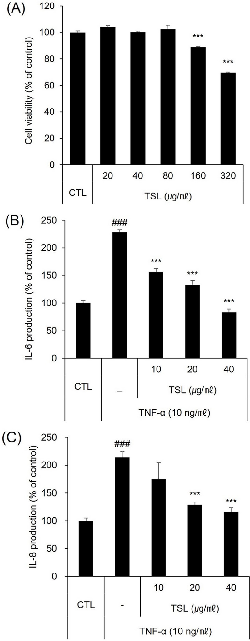
Inhibitory effects of TSL on inflammatory cytokine production in TNF-α–stimulated HaCaT cells.(A) Effects of TSL on HaCaT cell viability. (B, C) IL-6 and IL-8 expression, determined by ELISA. Data are presented as means ± SD. ***p < 0.001 vs. TNF-α alone; ###p < 0.001 vs. control (CTL).
These experiments showed that IL-6 and IL-8 production was increased in TNF-α–stimulated HaCaT cells; this effect was significantly attenuated by treatment with TSL (Fig. 1B and C). Thus, TSL decreased IL-6 and IL-8 expression at protein levels in TNF-α–stimulated HaCaT cells.
We also elucidated the anti-inflammatory effects of TSL in TNF-α-stimulated HS68 dermal fibroblasts. As shown in Fig. 2A, TSL was significantly cytotoxic at a concentration of 640 ㎍/㎖ in HS68 cells. To provide a suitable margin of safety, in subsequent experiments, we used a maximum TSL concentration of 160 ㎍/㎖.
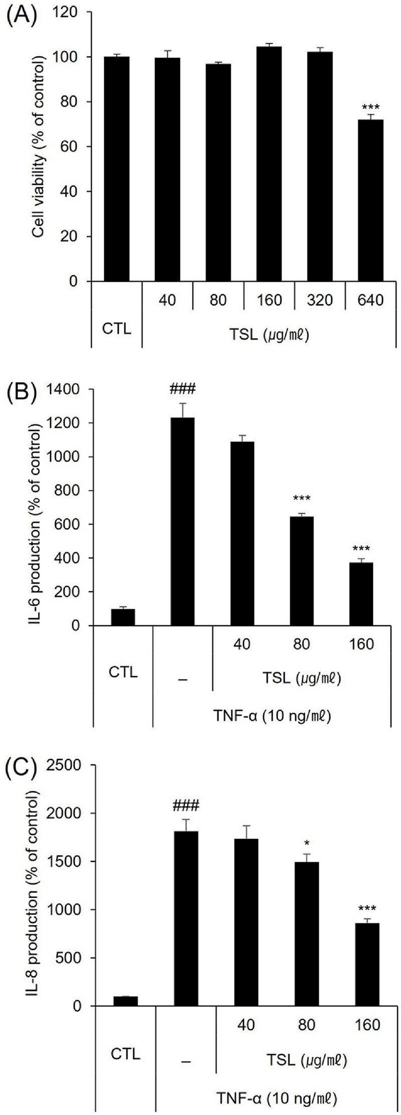
Inhibitory effects of TSL on inflammatory cytokine production in TNF-α-stimulated HS68 cells.(A) Effects of TSL on HS68 cell viability. (B, C) IL-6 and IL-8 expression, determined by ELISA. Data are presented as means ± SD. *p < 0.05, ***p < 0.001 vs. TNF-α alone; ###p < 0.001 vs. control (CTL).
Our investigation of the anti-inflammatory effect of TSL in TNF-α-stimulated HS68 cells using ELISA revealed that treatment with 40–160 ㎍/㎖ TSL caused a concentration-dependent reduction in IL-6 and IL-8 protein (Fig. 2B and C) levels in TNF-α–stimulated HS68 cells.
2. Inhibitory effect of TSL extract on MMP-1 production in TNF-α–stimulated HS68 dermal fibroblasts
To verify the anti-aging effects of TSL, we measured the production of MMP-1 at mRNA and protein levels using qRT-PCR and ELISA. TNF-α stimulation of HS68 cells increased MMP-1 mRNA and protein levels by 2.5 fold. Notably, TSL (40, 80, and 160 ㎍/㎖) significantly inhibited MMP-1 mRNA expression in TNF-α–stimulated HS68 cells (Fig. 3A).
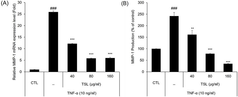
Inhibitory effect of TSL on MMP-1 production in TNF-α–stimulated HS68 cells.(A, B) MMP-1 expression, determined by qRT-PCR and ELISA. Expression of mRNA was normalized to that of GAPDH. Data are presented as means ± SD. **p < 0.01, ***p < 0.001 vs. TNF-α alone; ###p < 0.001 vs. controls (CTL).
Strikingly, at concentrations of 80 and 160 ㎍/㎖, TSL decreased secretion of MMP-1 protein to below control levels (Fig. 3B).
3. Antioxidant activity of TSL
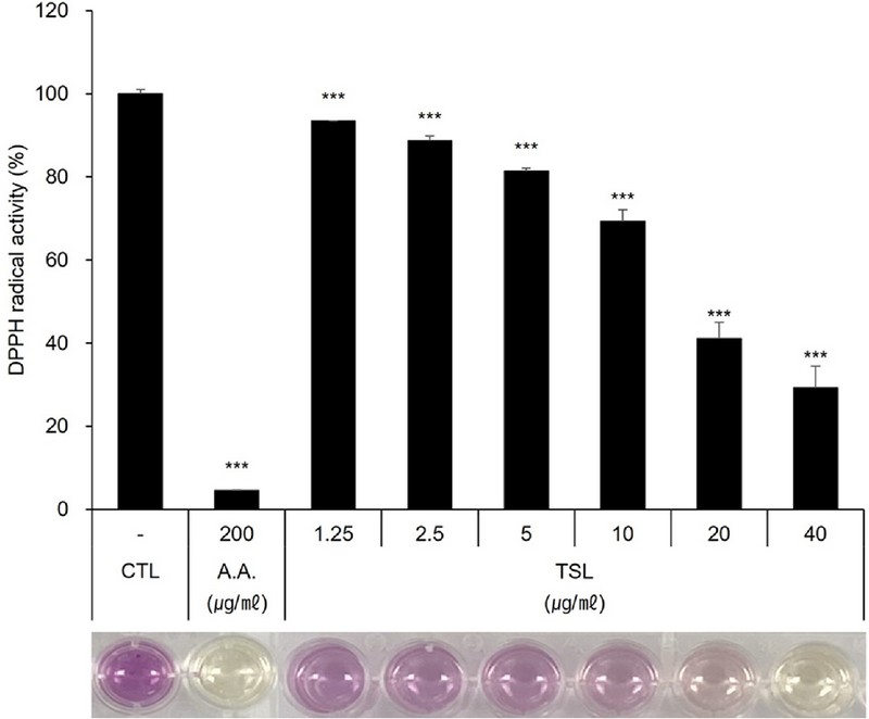
To examine the antioxidant effect of TSL, we conducted DPPH radical scavenging assays.
As shown in Fig. 4, TSL inhibited DPPH radical activity in concentration-dependent manner, decreasing it by up to 71.3% at a concentration of 40 ㎍/㎖. By comparison, 200 ㎍/㎖ ascorbic acid, used as a positive control, scavenged 96.2% of free radicals.
4. Anti-melanogenic effect of TSL on B16F10 melanoma cells
Finally, we assessed the anti-melanogenic efficacy of TSL in B16F10 cells, first performing cytotoxicity assays to identify a threshold concentration of TSL that affected cell viability.
We found that 40 ㎍/㎖ of TSL, the highest concentration tested, did not show cytotoxic effects on B16F10 cells (Fig. 5A). Accordingly, we treated α-MSH–stimulated B16F10 cells with 2.5, 5, 10, 20 and 40 ㎍/㎖ of TSL. As shown in Fig. 5B, α-MSH increased B16F10 cell secretion of melanin into culture media.
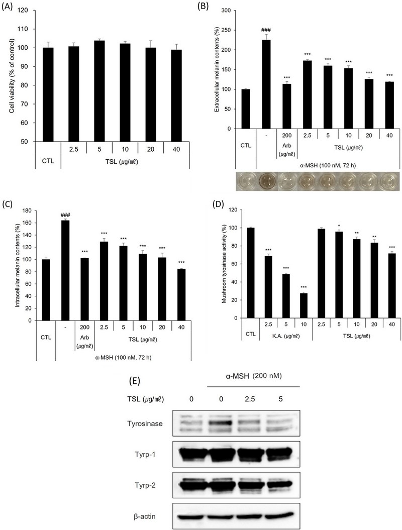
Anti-melanogenic effect of TSL. (A) Effects of TSL on B16F10 cell viability. (B, C) Effects of TSL on melanin content in B16F10 cells. Arbutin (Arb) was used as a positive control for assessment of melanin content in B16F10 cells. (D) Effects of TSL on mushroom tyrosinase activity. Kojic acid (K.A.) was used as a positive control for measurement of mushroom tyrosinase activity. Data are presented as means ± SD. *p < 0.05, **p < 0.01, ***p < 0.001 vs. α-MSH alone (B, C) or controls (D); ###p < 0.001 vs. controls (CTL). (E) Effects of TSL on the protein levels of melanogenic enzymes in B16F10 cells. β-actin was used as a loading control.
This effect was dramatically suppressed by TSL, which caused a concentration-dependent decrease in melanin secretion. TSL treatment also clearly inhibited intracellular melanin content in α-MSH–stimulated B16F10 cells in a concentration-dependent manner (Fig. 5C).
Arbutin, used as a positive control, inhibited both melanin production (intracellular levels) and secretion (extracellular levels). In addition, we determined the inhibitory efficacy of TSL on tyrosinase activity using mushroom tyrosinase activity assays, as described in methods.
These experiments showed that TSL significantly inhibited mushroom tyrosinase activity in a concentration-dependent manner (Fig. 5D). Kojic acid, used as positive control, also reduced the activity of mushroom tyrosinase.
Next, we performed western blotting assays to investigate the molecular mechanism on the anti-melanogenesis effect of TSL in α-MSH–stimulated B16F10 cells. In Fig. 5E, the protein levels of tyrosinase, Tyrp-1 and Tyrp-2 are increased by α-MSH in B16F10 cells. Data also showed that TSL reduced the protein expression of tyrosinase, Tyrp-1, and Tyrp-2 in α-MSH-stimulated B16F10 cells in a dose-dependent manner.
DISCUSSION
Generally speaking, the desirable attributes of skin treatments from a cosmetic perspective include anti-aging, antioxidant and anti-melanogenesis activities. Previous studies have established that extracts of Toona Sinensis possess a variety of desirable properties, including anti-inflammatory, anticancer, and antiviral effects (Peng et al., 2019; Chen et al., 2022).
However, not much is currently known about the pharmacological effects of Toona Sinensis on skin cells. Considering that TSL contains gallic acid, limonoids, and quercetin, which are preferred as cosmetic and nutritional ingredients, we here sought to study the potential cosmetic and pharmacological benefits that might be expected from application of TSL to skin cells.
In this study, we provide the first demonstration that TSL exerts anti-inflammatory, antioxidant, anti-melanogenesis, and anti-aging effects on appropriate in vitro cell models. IL-6 and IL-8 – representative pro-inflammatory cytokines – mediate skin inflammation caused by skin irritation (Tang et al., 2017).
In addition, induction of IL-6 and IL-8 by TNF-α is usually regulated by NF-κB (nuclear factor kappa-light-chain-enhancer of activated B cells) or MAPK (mitogen-activated protein kinase) signaling pathways (Grund et al., 2008; Lin et al., 2014).
We confirmed that TSL clearly inhibited IL-6 and IL-8 production in TNF-α–stimulated keratinocytes and dermal fibroblasts in this study, although additional studies will be required to determine whether the underlying molecular mechanisms involve inhibition of NF-κB or MAPK signaling pathway.
In addition, we assessed the anti-aging actions of TSL by investigating its effects on the expression level of MMP-1. Collagen, a major family of ECM proteins that plays a role in maintaining the tissue structure of the dermis, is degraded by MMP-1 in human aging skin (Järveläinen et al., 2009). Although TSL did not directly increase the expression of procollagen type I (data not shown), we found that it did significantly inhibit MMP-1 expression, indicating that TSL could exert an anti-aging effect on dermal fibroblasts through the suppression of collagen degradation.
We also confirmed the antioxidant effect of TSL in a cell-free system using DPPH scavenging assay. TSL showed antioxidant activity in all concentrations used in the experiment, and this result may be due to the presence of gallic acid, limonoids, and quercetin, which are known as antioxidants, in TSL. Future studies will determine whether TSL has antioxidant effects on skin cells. In addition, because enzymes such as catalase (CAT) and superoxide dismutase (SOD) are involved in antioxidant mechanisms (Sadi et al., 2008), we plan to test whether TSL increases the expression of these enzymes in skin cells as a measure of its antioxidant effects.
Lastly, we demonstrated that TSL significantly reduced α-MSH–stimulated increases in melanin in vitro. Tyrosinase plays a key role in promoting melanin synthesis during melano-genesis through two consecutive reactions: hydroxylation of L-tyrosine to L-DOPA, and oxidation of L-DOPA to DOPA-quinone, leading to skin pigmentation.
In the present study, we demonstrated that TSL clearly inhibited tyrosinase activity, suggesting that TSL could exert anti-melanogenic effects through tyrosinase inactivation. Thus, we evaluated the effect of CSL treatment on tyrosinase and its sub-signaling gene expression.
Two additional important contributors to melanogenesis are the transcription factor microphthalmia-associated transcription factor (MITF) and melanosomal enzyme tyrosinase related protein 1/2 (Tyrp-1/2) (Yasumoto et al., 1997; Bertolotto et al., 1998). Therefore, it is necessary to determine whether the anti-melanogenesis efficacy of TSL reflects regulation of MITF and/or Tyrp-1/2 protein expression. We confirmed by western blotting assay that TSL reduced the protein expression of tyrosinase, Tyrp-1, and Tyrp-2 in α-MSH–stimulated B16F10 cells.
Collectively, our findings confirmed several positive effects of TSL in skin physiology that suggest its potential pharmacological efficacy. Using TSL as an active ingredient in cosmetics will likely require demonstrating other beneficial effects of TSL, such as anti-acne and hair growth activity, in skin cells. Interesting in this context, TSL is known to exert antibacterial effects, inhibiting the acne bacterium P. acnes (Lim et al., 2020).
However, further experiments are needed to confirm these anti-acne effects such as sebum reduction at the cell level through Oil Red O staining or Nile Red staining using sebocytes. Moreover, the proliferative or protective effect of TSL on human hair follicle dermal papilla cells (HFDPCs) should be confirmed to establish the effect of TSL on hair growth.
It will also be important to investigate the expression of hair growth-promoting factors, such as vascular endothelial growth factor (VEGF), insulin-like growth factor (IGF-1), hepatocyte growth factor (HGF), and epidermal growth factor (EGF) (Choi, 2020; Zhou et al., 2021).
In conclusion, using the experimental findings presented here as a framework, we plan to further explore the potential of TSL as a cosmetic ingredient for anti-acne and anti-alopecia in a future study.
Collectively, our research suggests the novel possibility that TSL could be an efficacious skin treatment agent for further development in cosmetics and pharmaceuticals.
References
-
Ågren MS, Schnabel R, Christensen LH and Mirastschijski U. (2015). Tumor necrosis factor-α-accelerated degradation of type I collagen in human skin is associated with elevated matrix metalloproteinase(MMP)-1 and MMP-3 ex vivo. European Journal of Cell Biology. 94:12-21.
[https://doi.org/10.1016/j.ejcb.2014.10.001]

-
Bertolotto C, Buscà R, Abbe P, Bille K, Aberdam E, Ortonne JP and Ballotti R. (1998). Different cis-acting elements are involved in the regulation of TRP1 and TRP2 promoter activities by cyclic AMP: pivotal role of M boxes (GTCATGTGCT) and of microphthalmia. Molecular and Cellular Biology. 18:694-702.
[https://doi.org/10.1128/MCB.18.2.694]

-
Blanpain C and Fuchs E. (2006). Epidermal stem cells of the skin. Annual Review of Cell and Developmental Biology. 22:339-373.
[https://doi.org/10.1146/annurev.cellbio.22.010305.104357]

-
Chen CJ, Michaelis M, Hsu HK, Tsai CC, Yang KD, Wu YC, Cinatl J Jr and Doerr HW. (2008). Toona sinensis Roem tender leaf extract inhibits SARS coronavirus replication. Journal of Ethnopharmacology. 120:108-111.
[https://doi.org/10.1016/j.jep.2008.07.048]

-
Chen Y, Gao H, Liu X, Zhou J, Jiang Y, Wang F, Wang R and Li W. (2022). Terpenoids from the seeds of Toona sinensis and their ability to attenuate high glucose-induced oxidative stress and inflammation in rat glomerular mesangial cells. Molecules. 27:5784. https://www.mdpi.com/1420-3049/27/18/5784, (cited by 2024 February 8).
[https://doi.org/10.3390/molecules27185784]

-
Chia YC, Rajbanshi R, Calhoun C and Chiu RH. (2010). Anti-neoplastic effects of gallic acid, a major component of Toona sinensis leaf extract, on oral squamous carcinoma cells. Molecules. 15:8377-8389. https://www.mdpi.com/1420-3049/15/11/8377, (cited by 2024 June 19).
[https://doi.org/10.3390/molecules15118377]

-
Choi BY. (2020). Targeting Wnt/β-catenin pathway for developing therapies for hair loss. International Journal of Molecular Sciences. 21:4915. https://www.mdpi.com/1422-0067/21/14/4915, (cited by 2024 March 28).
[https://doi.org/10.3390/ijms21144915]

-
Dinarello CA. (1997). Interleukin-1. Cytokine and Growth Factor Reviews. 8:253-265.
[https://doi.org/10.1016/S1359-6101(97)00023-3]

-
Dong XJ, Zhu YF, Bao GH, Hu FL and Qin GW. (2013). New limonoids and a dihydrobenzofuran norlignan from the roots of Toona sinensis. Molecules. 18:2840-2850.
[https://doi.org/10.3390/molecules18032840]

-
D'Orazio J, Jarrett S, Amaro-Ortiz A and Scott T. (2013). UV radiation and the skin. International Journal of Molecular Sciences. 14:12222-2248.
[https://doi.org/10.3390/ijms140612222]

-
Fields GB. (2013). Interstitial collagen catabolism. Journal of Biological Chemistry. 288:8785-8793.
[https://doi.org/10.1074/jbc.R113.451211]

-
Grund EM, Kagan D, Tran CA, Zeitvogel A, Starzinski-Powitz A, Nataraja S and Palmer SS. (2008). Tumor necrosis factor-alpha regulates inflammatory and mesenchymal responses via mitogen-activated protein kinase kinase, p38, and nuclear factor kappaB in human endometriotic epithelial cells. Molecular Pharmacology. 73:1394-1404.
[https://doi.org/10.1124/mol.107.042176]

-
Hu J, Song Y, Mao X, Wang ZJ and Zhao QJ. (2016). Limonoids isolated from Toona sinensis and their radical scavenging, anti-inflammatory and cytotoxic activities. Journal of Functional Foods. 20:1-9.
[https://doi.org/10.1016/j.jff.2015.10.009]

-
Järveläinen H, Sainio A, Koulu M, Wight TN and Penttinen R. (2009). Extracellular matrix molecules: Potential targets in pharmacotherapy. Pharmacological Reviews. 61:198-223.
[https://doi.org/10.1124/pr.109.001289]

-
Jegal J, Kim TY, Park NJ, Jo BG, Jo GA, Choi HS, Kim SN and Yang MH. (2021). Inhibitory effects of luteolin 7-methyl ether isolated from wikstroemia ganpi on Tnf-A/Ifn-Γ mixture-induced inflammation in human keratinocyte. Nutrients. 13:4387. https://www.mdpi.com/2072-6643/13/12/4387, (cited by 2024 February 8).
[https://doi.org/10.3390/nu13124387]

-
Jeong HR, Kim JM, Lee U, Kang JY, Park SK, Lee HL, Moon JH, Kim MJ, Go MJ and Heo HJ. (2022). Leaves of Cedrela sinensis attenuate chronic unpredictable mild stress-induced depression-like behavior via regulation of hormonal and inflammatory imbalance. Antioxidants. 11:2448. https://www.mdpi.com/2076-3921/11/12/2448, (cited by 2024 February 8).
[https://doi.org/10.3390/antiox11122448]

- Kim SY, Lee MH, Jo NR and Park SN. (2010). Antibaterial activity and skin moisturizing effect of Cedrela sinensis A. Juss shoots extracts. Journal of Society of Cosmetic Scientists of Korea. 36:315-321.
-
Lauer-Fields JL and Fields GB. (2002). Triple-helical peptide analysis of collagenolytic protease activity. Biological Chemistry. 383:1095-1105.
[https://doi.org/10.1515/BC.2002.118]

-
Lee CS, Bae IH, Han J, Choi GY, Hwang KH, Kim DH, Yeom MH, Park YH and Park M. (2014). Compound K inhibits MMP-1 expression through suppression of c-Src-dependent ERK activation in TNF-α-stimulated dermal fibroblast. Experimental Dermatology. 23:819-824.
[https://doi.org/10.1111/exd.12536]

-
Lee CS, Jang WH, Park M, Jung K, Baek HS, Joo YH, Park YH and Lim KM. (2013). A novel adamantyl benzylbenzamide derivative, AP736, suppresses melanogenesis through the inhibition of cAMP-PKA-CREB-activated microphthalmia-associated transcription factor and tyrosinase expression. Experimental Dermatology. 22:762-764.
[https://doi.org/10.1111/exd.12248]

-
Lim HJ, Park IS, Jie EY, Ahn WS, Kim SJ, Jeong SI, Yu KY, Kim SW and Jung CH. (2020). Anti-inflammatory activities of an extract of in vitro grown adventitious shoots of Toona sinensis in LPS-treated RAW264. 7 and Propionibacterium acnes-treated HaCaT cells. Plants (Basel). 9:1701. https://www.mdpi.com/2223-7747/9/12/1701, (cited by 2024 February 8).
[https://doi.org/10.3390/plants9121701]

-
Lin HC, Lin TH, Wu MY, Chiu YC, Tang CH, Hour MJ, Liou HC, Tu HJ, Yang RS and Fu WM. (2014). 5-Lipoxygenase inhibitors attenuate TNF-α-induced inflammation in human synovial fibroblasts. PLoS One. 9:e107890. https://journals.plos.org/plosone/article?id=10.1371/journal.pone.0107890, (cited by 2024 February 8).
[https://doi.org/10.1371/journal.pone.0107890]

-
Mehrad B and Standiford TJ. (1999). Role of cytokines in pulmonary antimicrobial host defense. Immunologic Research. 20:15-27.
[https://doi.org/10.1007/BF02786504]

-
Park K, Lee JH, Cho HC, Cho SY and Cho JW. (2010). Down-regulation of IL-6, IL-8, TNF-α and IL-1β by glucosamine in HaCaT cells, but not in the presence of TNF-α. Oncology Letters. 1:289-292.
[https://doi.org/10.3892/ol_00000051]

-
Peng W, Liu Y, Hu M, Zhang M, Yang J, Liang F, Huang Q and Wu C. (2019). Toona sinensis: a comprehensive review on its traditional usages, phytochemisty, pharmacology and toxicology. Revista Brasileira de Farmacognosia. 29:111-124.
[https://doi.org/10.1016/j.bjp.2018.07.009]

-
Sadi G, Yilmaz O and Güray T. (2008). Effect of vitamin C and lipoic acid on streptozotocin-induced diabetes gene expression: mRNA and protein expressions of Cu–Zn SOD and catalase. Molecular and Cellular Biochemistry. 309:109-116.
[https://doi.org/10.1007/s11010-007-9648-6]

-
Tada M, Kohno M and Niwano Y. (2014). Alleviation effect of arbutin on oxidative stress generated through tyrosinase reaction with L-tyrosine and L-DOPA. BMC Biochemistry. 15:23. https://link.springer.com/article/10.1186/1471-2091-15-23, (cited by 2024 March 13).
[https://doi.org/10.1186/1471-2091-15-23]

-
Tang SC, Liao PY, Hung SJ, Ge JS, Chen SM, Lai JC, Hsiao YP and Yang JH. (2017). Topical application of glycolic acid suppresses the UVB induced IL-6, IL-8, MCP-1 and COX-2 inflammation by modulating NF-κB signaling pathway in keratinocytes and mice skin. Journal of Dermatological Science. 86:238-248.
[https://doi.org/10.1016/j.jdermsci.2017.03.004]

-
Wang KJ, Yang CR and Zhang YJ. (2007). Phenolic antioxidants from Chinese toon(fresh young leaves and shoots of Toona sinensis). Food Chemistry. 101:365-371.
[https://doi.org/10.1016/j.foodchem.2006.01.044]

-
Yang CJ, Huang YJ, Wang CY, Wang PH, Hsu HK, Tsai MJ, Chen YC, Bharath Kumar V, Huang MS and Weng CF. (2010). Antiproliferative effect of Toona sinensis leaf extract on non–small-cell lung cancer. Translational Research. 155:305-314.
[https://doi.org/10.1016/j.trsl.2010.03.002]

-
Yasumoto KI, Yokoyama K, Takahashi K, Tomita Y and Shibahara S. (1997). Functional analysis of microphthalmia-associated transcription factor in pigment cell-specific transcription of the human tyrosinase family genes. Journal of Biological Chemistry. 272:50-3509.
[https://doi.org/10.1074/jbc.272.1.503]

-
Zhang Y, Dong H, Wang M and Zhang J. (2016). Quercetin isolated from Toona sinensis leaves attenuates hyperglycemia and protects hepatocytes in high-carbohydrate/high-fat diet and alloxan induced experimental diabetic Mice. Journal of Diabetes Research. 2016:8492780.
[https://doi.org/10.1155/2016/8492780]

-
Zhou Y, Liu Q, Bai Y, Yang K, Ye Y, Wu K, Huang J, Zhang Y, Zhang X, Thianthanyakij T, Wang J, Zhu Y, Lin J and Wu W. (2021). Autologous activated platelet-rich plasma in hair growth: a pilot study in male androgenetic alopecia with in vitro bioactivity investigation. Journal of Cosmetic Dermatology. 20:1221-1230.
[https://doi.org/10.1111/jocd.13709]

