
아까시나무와 약재 자괴화(콩과)의 화피 및 화분 미세형태학적 연구
This is an open access article distributed under the terms of the Creative Commons Attribution Non-Commercial License (http://creativecommons.org/licenses/by-nc/3.0/) which permits unrestricted non-commercial use, distribution, and reproduction in any medium, provided the original work is properly cited.
Abstract
Robinia pseudoacacia L. and its herbal product, Robiniae Flos, are traditionally used in Korea. However, commercially processed floral materials often exhibit similar morphological characteristics despite originating from different botanical sources. This study aimed to establish reliable authentication criteria by comparing the micromorphologies of fresh R. pseudoacacia flowers and Robiniae Flos.
Floral structures (petals, sepals, and pollen) were examined using stereo, light, and field-emission scanning electron microscopy. Petal epidermal features, including papillose and tabular flat cells and densely pubescent sepals, were identified. The pollen grains were monads and tricolporate with psilate–perforate ornamentation. No significant micromorphological differences were observed between the fresh and herbal samples. However, the pollen of the potentially confusable species, Styphnolobium japonicum, was significantly smaller, enabling clear discrimination between R. pseudoacacia and Robiniae Flos.
Fresh and commercial samples of R. pseudoacacia exhibited highly similar micromorphological traits. However, pollen samples from S. japonicum were distinct, providing a basis for discrimination. These findings provide reliable micromorphological criteria for distinguishing Robiniae Flos from other herbal products of morphologically similar species.
Keywords:
Authentication, Field Emission Scanning Electron Microscope, Pollen, Robinia pseudoacacia L., Robiniae Flos서 언
콩과 (Fabaceae = Leguminosae)는 약 750여 속, 19,000여 종으로 구성된 대규모 식물군으로, 전 세계에 널리 분포한다 (Polhill and Raven, 1981; Lewis et al., 2005; Xu and Deng, 2017). 한반도에는 최근 콩과의 분류체계에 따라 박태기나무아과 (Cercidoideae Legume Phylogeny Working Group), 실거리나무아과 (Caesalpinioideae DC.), 콩아과 (Papilionoideae DC.)로 총 3개 아과 (subfamily)에 속하는 48속 122종이 분포한다 (Choi, 2024).
이 중 실거리나무아과에 속하는 결명자속 (Senna Mill.), 자귀나무속 (Albizia Durazz.), 콩아과에 속하는 감초속 (Glycyrrhiza L.), 강낭콩속 (Phaseolus L.), 개자리속 (Medicago L.), 고삼속 (Sophora L.), 동부속 (Vigna Savi), 콩속 (Glycine Willd.), 황기속 (Astragalus L.), 회화나무속 (Styphnolobium Schott) 등에 속하는 식물은 식용, 약용 및 원예 자원으로서 높은 활용 가치를 지닌다 (Chung et al., 1998; Korea Institute of Oriental Medicine, 2026). 특히 콩아과 아까시나무속 (Robinia L.)에 속하는 아까시나무 (R. pseudoacacia L.)는 북아메리카 원산으로, 토양 및 수분 보전과 생태복원 효과가 우수한 종으로 알려져 있으며 (Vitkova et al., 2017), 국내에서는 주요 조림수종으로 널리 식재되어 왔다. 또한 밀원식물로서의 가치 역시 높은 것으로 보고되고 있다 (Hanover, 1992; Han et al., 2009; Zhu and Turland, 2010).
한편, 아까시나무의 건조된 꽃은 전통적으로 자괴화 (刺槐花, 생약명: Robiniae Flos)로 사용되어 왔으며, 조선민주주의인민공화국 약전 제8판에는 그 기원식물로 수재되어 있다 (Pharmacopoeia Commission of the Democratic People’s Republic of Korea, 2018). 특히 자괴화는 전통적으로 이뇨 (diuretic), 진경제 (spasmolytic) 및 담즙 분비 촉진 (cholagogic agents) 작용을 가진 것으로 알려져, 신장 (kidneys) 및 담도 (biliary ducts)의 염증 완화 (relieve inflammation)에 사용되어 왔다 (Pietta et al., 1998; Strzelecka and Kowalski, 2000). 최근 연구에서는 아까시나무 꽃에 다양한 플라보노이드 (flavonoid) 계열 화합물이 함유되어 있음이 보고되었으며 (Veitch et al., 2010), 탄닌 (tannin) 및 페놀성 화합물 (phenolic compounds)이 풍부하여 우수한 항산화 (antioxidant) 활성을 나타내는 것으로 확인되었다 (Stankov et al., 2018; Bratu et al., 2021). 또한, 항응고 (anticoagulant), α-glucosidase 저해를 포함한 다양한 생리활성 (Han et al., 2022)과 함께 미생물의 생물막 형성 억제 (antibiofilm) 및 군집감지 저해 (anti-quorum sensing) 활성을 나타내어 미생물 생장 억제와 군집행동 조절 측면에서의 약리적 활용 가능성이 제시되었다 (Uzelac et al., 2023).
자괴화는 꽃의 외형이 유사한 콩과 식물과 혼입 (adulteration)될 가능성이 있으나, 이에 대한 형태학적 감별 기준 및 미세형태 기반 연구는 상대적으로 부족한 실정이다. 실제로 콩과 식물 중 꽃을 약용하는 종으로 회화나무 [Styphnolobium japonicum (L.) Schott ≡ Sophora japonica L.]의 꽃인 괴화 (槐花, 생약명: Sophorae Flos)는 유통 과정에서 자괴화와 혼입되어 존재하는 사례가 보고된 바 있다 (Wei et al., 2019). 두 분류군은 개화 시기나 열매 형태에서는 차이를 보이나, 꽃의 형태가 유사하여 (Korea National Arboretum, 2016; Choi, 2024), 혼입 가능성이 높은 것으로 알려져 있다.
또한 콩과 식물뿐만 아니라, 약재 상태에서는 형태적으로 유사한 근연 분류군 간 혼입 및 대체 유통 사례가 빈번하게 발생하고 있으며, 건조 약재의 경우 외형이 매우 유사하여 정확한 기원 식물의 판별이 어려운 것으로 보고되었다 (Han et al., 2011; Lu et al., 2017; Zhang et al., 2019). 이러한 혼입은 약재의 품질 저하 및 안전성 문제로 이어질 수 있어 (Choudhary, 2021), 형태학적 기준에 기반한 감별 지표의 확립이 요구된다. 특히 임상에서의 독성 사고 예방과 품질 관리를 위해 다양한 기법을 활용한 약재의 정확한 감별과 기원종 동정의 중요성이 더욱 강조되고 있다 (World Health Organization, 2011; Newmaster et al., 2013; Chen et al., 2014; Zhu et al., 2022).
콩과 식물의 꽃 미세형태학적 특성은 종 수준에서 안정적으로 유지되는 형질로 알려져 있으며, 분류학적으로 중요한 지표로 활용되어 왔다 (Çildir et al., 2012). 특히 화판 (꽃잎, petals)과 악편 (꽃받침, sepals)의 표피세포 (epidermal cells) 구조, 모용 (trichome), 기공 복합체 구조 (stomatal complex), 그리고 화분 입자 (pollen grains) 형태와 같은 미세형태학적 형질은 종간 형태적 차이를 반영하는 주요 특징으로 보고된다 (Grohar et al., 2021). 이러한 형질들은 다양한 콩과 분류군에서 종 동정 및 분류를 위한 진단 형질, 바이오마커 (biomarker)로 활용될 수 있음이 입증되었다 (Kahraman et al., 2014; Liao et al., 2022; Khan et al., 2025). 한편, 화판을 포함한 화피의 미세형태학적 형질은 건조 약재 시료에서도 조직 연화 (tissue softening) 및 임계점 건조 (critical point drying) 전처리 과정을 통해 안정적으로 관찰 가능하여, 최근 약재 감별을 위한 유효한 형질로 주목받고 있다 (Serrano et al., 2010; Konarska et al., 2023; Obika and Obika, 2023; Lee et al., 2025).
따라서 본 연구에서는 실체현미경 (Stereomicroscope, SM), 광학현미경 (Light microscopy, LM) 및 주사전자현미경 (Scanning electron microscopy, SEM)을 이용하여, 아까시나무 꽃과 자괴화 유통 약재의 미세형태학적 특성을 상세히 관찰하고, 이를 상호 비교·분석함으로써 명확한 감별 기준을 제시하고자 하였다. 또한 꽃의 외부 형태가 유사하여 혼입 가능성이 있는 회화나무와의 명확한 분류 기준을 확립하기 위해, 약재 상태에서도 확인 가능한 화분의 미세형태학적 비교 분석을 수행하였다.
재료 및 방법
1. 연구 재료
아까시나무의 꽃은 2025년 5월 개화기에 충청북도 청주시에서 채집하였으며, 확보한 단일 개체는 70% 에탄올로 고정하여 액침표본으로 제작하고, 일부는 석엽표본으로 제작하여 보관하였다 [Robinia pseudoacacia; Korea, Chungcheongbuk-do, Cheongju-si, 7. May. 2025. Lee 612 (CBU, herbarium acronym)]. 약재 자괴화는 국내 온라인 한약재 시장에서 구입한 건조시료를 실험에 사용하였다 [Robiniae Flos; Korea, Chungcheongbuk-do, Chungju-si, 2025. 544 (CBU)]. 또한 화분학적 비교를 위해 회화나무 석엽표본으로부터 화부 기관을 채취하여 사용하였다 [Styphnolobium japonicum; Korea, Chungcheongbuk-do, Cheongju-si, 12. Jul. 2024. JI 256 (CBU)].
2. 외부 형태학적 형질
아까시나무 생화의 생태사진은 디지털카메라 (digital camera; Samsung WB100, Samsung Electronics, Suwon, Korea)를 이용하여 촬영하였다. 생식기관 및 외부 형태는 실체현미경 (SM; S9D, Leica, Wetzlar, Germany)을 이용하여 관찰하고 측정하였다.
3. 화판 및 악편 미세형태학적 형질
화피 (화판, 악편)의 미세형태학적 형질 관찰을 위해 채집된 아까시나무 액침 시료를 임계점 건조기 (critical point dryer; SPI-13200J-AB, SPI Supplies, West Chester, PA, USA)를 이용하여 완전 건조하였다. 건조된 시료는 직경 9.5 mm 알루미늄 스터브 (aluminum stub; 01501-BA, SPI Supplies, West Chester, PA, USA)에 카본 테이프 (carbon tape; RS-MN-15-000412, SPI Supplies, West Chester, PA, USA)로 고정한 후, 이온증착기 (ion sputter coater; Q150T ES Plus, Quorum, UK)를 사용하여 백금 (Pt)으로 15초간 코팅하였다. 이후 주사전자현미경 (SEM; EM-30, COXEM, Daejeon, Korea)을 이용하여 가속전압 5 kV, 작동거리 5–7 mm 조건에서 관찰하였다.
건조 자괴화 약재 시료는 약 90°C에서 30분간 열탕 처리한 후, 연화제 (softening agent; Agepon, Agfa-Gevaert, Leverkusen, Germany)와 증류수를 배합한 용액 (Agepon : 증류수 = 1 : 200)에 12시간 이상 침지하여 조직을 연화시켰다. 이후 70% 에탄올에 보관된 시료는 에탄올 시리즈 (80%, 90%, 100%)를 따라 각각 15분씩 탈수하였다. 탈수된 시료는 아이소아밀 아세테이트 (isoamyl acetate; Daejung Chemicals & Metals Co., Korea)에 15분 이상 치환한 후, 임계점 건조기를 이용하여 완전 건조하였다. 건조된 시료는 생체 시료와 동일한 조건 하에서 검경하였다.
4. 화분의 미세형태
화분 형태를 관찰하기 위해 아까시나무 생화 및 자괴화 약재 시료, 그리고 회화나무의 건조된 약 (anther)을 루골 용액 (Lugol’s solution)으로 3분간 염색한 후 광학현미경 (LM; DM750, Leica, Wetzlar, Germany)을 이용하여 관찰하였다. 루골 용액은 화분 내부의 세포질 (특히 전분)을 염색하여 전체 형태를 확인하기 위해 사용하였다.
약에서 화분을 방출시켜 칼베를라 용액 (Calberla’s solution)으로 5분 이상 염색한 후 동일한 광학현미경으로 관찰하였다. 칼베를라 용액은 화분의 팽윤을 유도하고 형태 변형을 최소화하여 정량적 분석의 정확도를 향상시키기 위해 사용하였다. 단일 개체 각 시료에서 염색된 화분 30개를 무작위로 선별하여 극축 길이 (polar axis length, P)와 적도 직경 (equatorial diameter, E)을 측정하였다. 아까시나무 생화와 자괴화의 화분 미세형태는 각 시료에서 채취한 약을 임계점 건조기를 이용하여 건조한 후, 고배율·고해상도 이미지를 확보하기 위해 전계방출형 주사전자현미경 (Field Emission-SEM; Ultra Plus, Carl Zeiss, Jena, Germany)을 이용하여 화분 외벽의 표면 무늬를 관찰하였다. 형질에 대한 용어는 Punt et al. (2007)을 따랐다.
5. 통계 분석
연구 결과는 평균 ± 표준편차 (mean ± standard deviation)로 나타내었다. 아까시나무 생화, 자괴화 및 회화나무의 화분에서 관찰된 정량적 형질 (P, E 및 P/E 비율)의 그룹 간 차이를 비교하기 위해 일원분산분석 (one-way analysis of variance, ANOVA)을 수행하였으며, 사후검정으로 Tukey의 다중비교검정 (Tukey’s honestly significant difference test)을 실시하였다. 모든 통계 분석은 R 통계 프로그램 (R version 4.5.2, R Foundation for Statistical Computing, Vienna, Austria)을 사용하였다. 통계적 유의성은 P < 0.001 수준에서 검증하였다.
결 과
1. 꽃의 외부 형태학적 형질
아까시나무 (R. pseudoacacia)의 꽃과 자괴화 (Robiniae Flos)의 외부 형태를 해부하여 관찰한 결과, 두 시료 모두 동일한 구조를 나타냈다. 화서 (inflorescence)는 액생 (axillary)하는 총상화서 (raceme)이다 (Fig. 1A-B). 꽃은 양성화 (bisexual)이고, 좌우대칭 (bilaterally symmetrical)의 접형화관 (papilionaceous corolla)이며 (Fig. 1C), 악 (calyx)은 종형 (campanulate)으로 악편 (calyx lobes)은 5열 (5-lobed)한다 (Fig. 1D). 화판 (petals)은 백색으로 기판 (standard = banner) 내부에는 황색 반점 (yellow spot)이 있고, 아원형 (suborbicular)이며, 반곡하고 (reflexed), 선단은 미요두 (retuse)이다 (Fig. 1E, I, J). 익판 (wings)은 좁은 도란형 (narrowly obovate)이고, 선단은 원두 (rounded)이며 (Fig. 1F, K), 용골판 (keels)은 넓은 삼각형 (broadly triangular)이고, 선단은 예두 (acute)이다 (Fig. 1G, L). 수술 (stamens)은 양체웅예 (diadelphous)이고, 10 (9+1)개로 구성되며, 한 개의 수술은 이생 (free)하고, 나머지 9개 수술은 서로 유합 (connate)한다 (Fig. 1H, M). 암술 (pistil)은 1개이며, 자방 (ovary)에는 자루 (stipe)가 있고, 화주 (style)는 만곡 (incurved)하며, 배주는 다수이다.
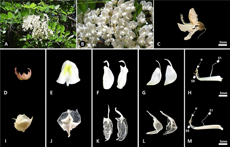
Photographs of R. pseudoacacia and Robiniae Flos.A-B; Habit, C; Robiniae Flos, D-H; Dissected floral organs of R. pseudoacacia, I-M; Dissected floral organs of Robiniae Flos, D; Calyx, E, I, J; Standard, F, K; Wings, G, L; Keels, H, M; Stamens and pistil. P; Pistil, S1; One stamen of unequal size, S9; Nine stamens of unequal size.
2. 화판 표피의 미세형태학적 형질
아까시나무의 꽃과 자괴화의 화판 표피 구조는 기판, 익판 및 용골판의 외면 (abaxial = outer side of petal)과 내면 (adaxial = inner side of petal)으로 구분하여 관찰하였다. 화판 표피의 수층벽 (anticlinal wall)은 외면과 내면 모두에서 직선형 (straight)과 곡선형 (curved)이 혼재된 형태로 나타났으며, 특히 외면에서는 곡선형과 파상형 (undulate)이 함께 관찰되었다. 병층벽 (periclinal wall)은 외면과 내면 모두에서 유선상 줄무늬 (striation)가 확인되었다 (Fig. 2; Table 1). 표피세포의 형태는 등방형 (isodiametric)과 불규칙형 (irregular)이 혼재하였으며, 기공과 모용은 관찰되지 않았다.
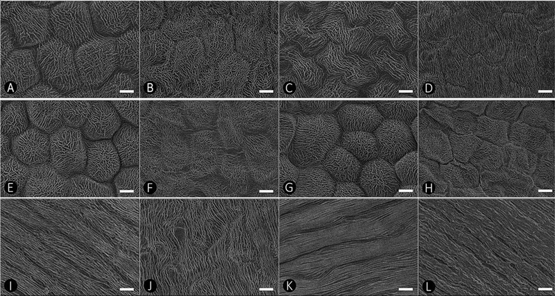
Scanning electron microscopy images of petal epidermis in R. pseudoacacia and Robiniae Flos.A, C, E, G, I, K; R. pseudoacacia, B, D, F, H, J, L; Robiniae Flos, A-D; Standard petal, E-H; Wing petals, I-L; Keel petals, A-B, E-F, I-J; Outer side, C-D, G-H, K-L; Inner side. Scale bars = 10 μm.

Comparison of petal micromorphological characteristics between R. pseudoacacia and Robiniae Flos.
화판 표피세포는 주름진 유두상 혹형 세포 (PKR, papillose knobby cells with a rugose sculpture)와 줄무늬 판상 편평형 세포 (TFS, tabular flat cells with longitudinal striations)의 두 유형으로 구분되었다 (Fig. 2; Table 1).
아까시나무 꽃과 자괴화의 기판 외면과 내면에서는 주름진 유두상 혹형 세포 (PKR)가 관찰되었으며, 익판의 외면과 내면에서도 두 시료 모두 동일한 세포 유형이 나타났다. 반면, 용골판의 외면과 내면에서는 두 분류군 모두 줄무늬 판상 편평형 세포 (TFS)가 관찰되었다 (Fig. 2).
3. 악편 표피의 미세형태학적 형질
아까시나무 꽃과 자괴화 모두에서 악편 표면에 단세포 단모 (unicellular non-glandular trichome)가 밀집하여 분포 (densely distributed)하고 있어 표피 구조를 관찰하기 어려웠다 (Fig. 3A-B). 반면, 화판에서는 관찰되지 않았던 기공이 확인되었으며, 기공 복합체는 부재형 (anomocytic type)으로 나타났다 (Fig. 3C-D).
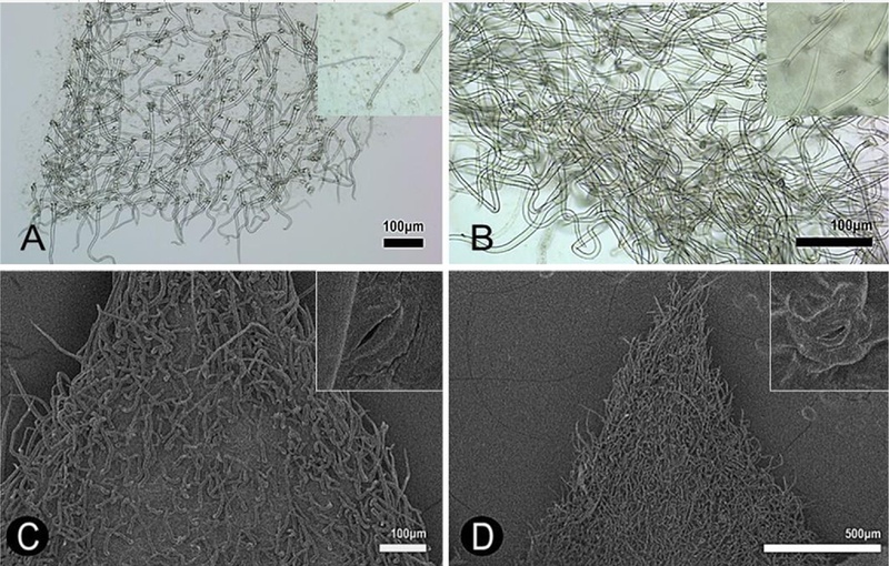
Light and scanning electron microscopy images of sepals in R. pseudoacacia and Robiniae Flos.A, C; R. pseudoacacia, B, D; Robiniae Flos, A-B; LM images (inset: close-up of the trichome), C-D; SEM images (inset: close-up of the stomata complex). All images show the outer surface.
4. 화분의 미세형태학적 형질
아까시나무 생화와 약재 자괴화 시료의 화분은 모두 단립 (monad)으로 발아구는 삼공구형 (tricolporate)이고, 표면 무늬는 평활상 유공형 (psilate–perforate)으로 확인되었다 (Fig. 4A-D). 극축 길이는 아까시나무 생화에서 26.9 ± 1.15 ㎛, 자괴화에서 28.6 ± 1.98 ㎛로 나타났으며, 적도 직경은 각각 30.6 ± 1.58 ㎛와 32.9 ± 2.31 ㎛로 관찰되었다 (Table 2). 반면, 회화나무에서는 극축 길이와 적도 직경이 각각 15.5 ± 0.63 ㎛와 16.3 ± 0.60 ㎛로 나타나 유의하게 작게 확인되었다 (P < 0.001; Table 2). 또한, P/E값은 아까시나무에서 0.88 ± 0.06, 자괴화에서 0.87 ± 0.08, 회화나무에서 0.95 ± 0.05로 나타나, 모든 시료에서 약단구형 (oblate-spheroidal)이 우세하였다 (Table 2).
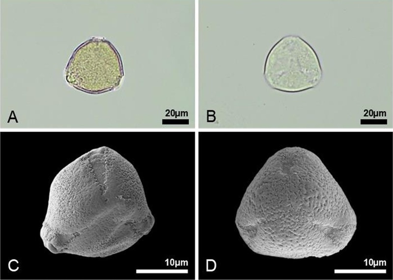
Light and scanning electron micrographs of pollen grains of R. pseudoacacia and Robiniae Flos in polar view.A, C; R. pseudoacacia, B, D; Robiniae Flos, A, B; LM images, C, D; SEM images.
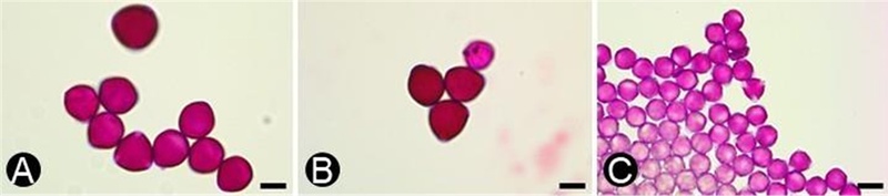
Light microscopy images of pollen grains of R. pseudoacacia, Robiniae Flos, and S. japonicum in polar view.A; R. pseudoacacia, B; Robiniae Flos, C; S. japonicum. Scale bars = 20 ㎛.
Measurements of pollen grains of R. pseudoacacia, Robiniae Flos, and S. japonicum.
고 찰
본 연구에서는 자괴화 (Robiniae Flos)와 그 기원종인 아까시나무 (Robinia pseudoacacia) 생화를 대상으로 화판, 악편 및 화분의 미세형태학적 형질을 비교·분석하였다. 또한 화부기관이 약재로 유통되는 경우 꽃 내부에 포함된 화분학적 형질이 감별 기준이 될 수 있는지에 대한 가능성을 확인하고자 자괴화와 동일 과 (family), 동일 기관 (organ)이 약재로 수재되어 있는 회화나무 (Styphnolobium japonicum) 생화 화분을 함께 비교하여 실질적인 약재 감별에 활용 가능한 형태학적 지표를 제시하고자 하였다.
본 연구에서는 정확한 종 동정을 거쳐 직접 채집한 아까시나무와 회화나무 시료, 그리고 유통 중인 약재 자괴화를 대상으로 하였다. 미세구조는 주사전자현미경을 이용하여 관찰하였으며, 일부 형질은 광학현미경 검경을 병행하여 분석하였다.
화판 표피의 유두상 세포 (papillose epidermal cells) 조합은 꿀벌, 나비 등 수분매개자 (pollinator)의 착지 안정성을 높이고 채밀 시 미끄럼을 방지하는 기능적 형질로 보고된 바 있다 (Kevan and Lane, 1985; Whitney et al., 2009; Córdoba and Cocucci, 2011; Aronne et al., 2012). 이러한 화판 구조는 수분매개자의 채집 효율 및 꽃 선택에 영향을 미치는 것으로 알려져 있다 (Giovanetti, 2019). 또한 꽃의 형태와 구조는 특정 수분매개자에 적합하도록 진화한 기능적 형질로서, 물리적 상호작용을 통해 수분 과정을 조절하는 역할을 수행하는 것으로 보고되었다 (Kobayashi et al., 2019). 본 연구에서도 아까시나무 생화와 자괴화는 화판 표피세포 유형의 분포 양상이 유사하게 나타났으며, 기판과 익판에서는 주름진 유두상 혹형 세포 (PKR)가, 용골판에서는 줄무늬 판상 편평형 세포 (TFS)가 일관되게 관찰되었다. 이는 두 시료가 동일한 기원종임을 시사하는 동시에, 채밀 과정에서 수분매개자가 기판과 익판 내면의 세포 구조에 의해 착지 안정성을 확보할 가능성을 나타낸다. 또한 이러한 결과는 콩과 식물에서 화판 표피 형질이 종 수준에서 안정적으로 유지된다는 선행 연구와 일치하며 (Ojeda et al., 2009; Çildir et al., 2012), 화판 표피 구조가 분류학적 진단 형질로 활용될 수 있음을 제시한 기존 연구를 지지한다 (Bailes and Glover, 2018; Shah et al., 2023; Badry et al., 2024).
악편 표피에서는 아까시나무와 자괴화 모두 단세포 단모가 밀집하여 분포하는 형태가 관찰되었다. 콩과 식물에서 악편의 모용은 화기관을 보호하고 외부 환경과의 상호작용에 관여하는 구조로 보고된 바 있다 (Vrachnakis et al., 2005). 또한 선행 연구에 따르면, 콩과 식물에서 모용은 형태학적 및 분류학적 진단 형질로 활용될 수 있으며 (Çildir et al., 2012), 특히 아까시나무속 (Robinia)에서 모용의 형태와 구조가 분류학적 구분에 유용한 형질로 보고된 바 있다 (Konarska and Łotocka, 2020). 본 연구 결과, 이러한 구조적 특징은 건조 및 가공 과정을 거친 약재 상태에서도 안정적으로 유지되는 형질로 확인되었으며, 이전 연구에서처럼 (Muyumba et al., 2021), 약재 감별을 위한 유용한 형태학적 지표로 활용될 수 있을 것으로 판단하였다.
약재 유통 과정에서는 형태적으로 유사한 꽃 약재 간 혼입 및 대체 사례가 지속적으로 보고되고 있으며 (Han et al., 2011; Lu et al., 2017; Wei et al., 2019; Zhang et al., 2019), 실제로 괴화 (Sophorae Flos) 유통 과정에서 자괴화와의 혼입 사례가 확인된 바 있다 (Wei et al., 2019). 이러한 문제는 약재의 품질 관리 및 안전성 측면에서 중요한 과제로 인식되고 있다. 본 연구에서 수행한 화분 형태 비교 결과, 세 시료 모두에서 삼공구형이 관찰되었다. 화분 크기 및 P/E 비율 비교에서는 아까시나무와 자괴화 간 유의한 차이가 나타나지 않은 반면, 회화나무에서는 통계적으로 유의한 차이가 확인되었다 (P < 0.001). 선행 연구에 따르면, 화분 크기와 형태는 건조 및 가공과정 이후에도 안정적으로 유지되는 형질로 보고되었다 (Govindarajan et al., 2019). 특히 콩과 식물에서 화분 미세형태는 종 수준 분류에 유용한 형질로 보고되어 왔으며 (Song, 2007), 다양한 분류군에서 공통적인 화분 유형이 확인되는 동시에 화분 크기 (P, E)와 표벽 구조 및 무늬 (exine ornamentation)의 차이에 의해 종 간 구분이 가능함이 제시되었다 (Bahadur et al., 2022; Liao et al., 2022; Khan et al., 2022; Das et al., 2025; Khan et al., 2025). 따라서 약용 부위가 꽃인 약재의 경우, 화분 크기 측정과 같은 간단한 검증을 통해 기원식물의 판별이 가능할 것으로 판단되며, 화분학적 형질은 약재 감별을 위한 신뢰성 있는 형태학적 지표로 활용될 수 있을 것으로 판단된다. 비록 본 결과는 화분학적 형질이 감별 기준으로 활용될 가능성을 확인한 수준에 해당하지만, 향후 화부 기관을 약재로 사용하는 품목들의 유통품 및 채집품을 폭넓게 수집하여 종합적으로 비교·검토함으로써, 보다 명확한 화분학적 감별 기준과 지표를 확립할 필요가 있을 것으로 판단된다.
전반적으로 화판의 표피세포 유형, 세포벽 구조, 악편의 모용 및 기공 복합체 유형, 그리고 화분의 형태와 표벽 무늬 등과 같은 질적 형질은 생화와 자괴화 간에 유의한 차이를 보이지 않았다. 이는 건조 및 가공 과정을 거친 약재 상태에서도 기원식물의 미세형태학적 특성이 안정적으로 유지됨을 시사한다. 실제로 건조된 식물 시료는 적절한 연화 및 전처리 과정을 통해 조직을 복원한 뒤 현미경 관찰이 가능하며, 이러한 미세형태학적 분석은 약재의 동일성 평가를 위한 표준적인 방법으로 활용되고 있다 (World Health Organization, 2011). 따라서 본 연구 결과는 건조 시료에서도 미세형태학적 형질을 활용한 감별이 가능함을 보고한 기존 연구들과 일치하며 (Govindarajan et al., 2019; Younus et al., 2019), 본 연구에서 확인된 형질 또한 실제 유통 약재의 감별에 적용 가능한 신뢰성 있는 지표로 활용될 수 있음을 시사한다.
본 연구는 미세형태학적 및 화분학적 형질을 활용하여 기원종과 유통 약재를 비교·분석함으로써, 참조 표준으로 활용 가능한 형태학적 감별 기준을 제시하였다는 점에서 의의가 있다. 또한 혼입 가능성이 높은 약재 기원종에 대해 화분학적 형질의 감별 적용 가능성을 확인하였다. 향후 연구에서는 다양한 지역과 개체군을 포함한 시료 확대와 함께, 형태적으로 유사한 종 및 화부를 약용 부위로 사용하는 생약을 추가적으로 비교하여 감별 기준의 범용성을 검증할 필요가 있다. 나아가 DNA 바코딩과 화학성분 분석을 결합한 다중 분석 기반의 표준화된 감별 프로토콜을 구축하고, 다빈도 약재를 중심으로 현장 적용이 가능한 자동화 판별 기술로의 확장을 고민해야 하겠다.
Acknowledgments
본 연구는 2026년도 식품의약품안전처의 연구개발비(25202한약안006)로 수행되었으며 이에 감사드립니다.
References
-
Aronne G, Giovanetti M and De Micco V. (2012). Morpho-functional traits and pollination mechanisms of Coronilla emerus L. flowers(Fabaceae). The Scientific World Journal. 2012:381575. (cited by 2026 Apr 26).
[https://doi.org/10.1100/2012/381575]

-
Badry MO, Osman AK, Aboulela M, Gafar S and Nour IH. (2024). Taxonomic implications of normal and abnormal stomatal complexes in Indigofera L.(Indigofereae, Faboideae, Fabaceae). Protoplasma. 261:991-1021.
[https://doi.org/10.1007/s00709-024-01951-0]
-
Bahadur S, Taj S, Long W and Hanif U. (2022). Pollen morphological peculiarities of selected Mimosoideae taxa of Hainan Island and their taxonomic relevance. Agronomy. 12:1122. (cited by 2026 Apr 26).
[https://doi.org/10.3390/agronomy12051122]
-
Bailes EJ and Glover BJ. (2018). Intraspecific variation in the petal epidermal cell morphology of Vicia faba L.(Fabaceae). Flora. 244:29-36.
[https://doi.org/10.1016/j.flora.2018.06.005]
-
Bratu MM, Moldovan R, Gardan IA, Voicu L and Gherman MC. (2021). Evaluation of the antioxidant, cytotoxic and antitumoral activities of a polyphenolic extract of Robinia pseudoacacia L. flowers. Journal of Science and Arts. 21:195-206.
[https://doi.org/10.46939/J.Sci.Arts-21.2-b04]
-
Chen S, Pang X, Song J, Shi L, Yao H, Han J and Leon C. (2014). A renaissance in herbal medicine identification: from morphology to DNA. Biotechnology Advances. 32:1237-1244.
[https://doi.org/10.1016/j.biotechadv.2014.07.004]
- Choi BH. (2024). Fabaceae Lindl. (Leguminosae Juss.) In Flora of Korea Editorial Committee (eds.). Flora of Korea. Volume 5a-3 (Rosidae: Fabaceae). Junghaengsa. Incheon, Republic of Korea. p.3-87.
- Choudhary JM. (2021). Introduction to adulteration of crude drug and its evaluation methods: concise review. International Journal of Pharmaceutical Research and Applications. 6:774-781.
-
Çildir H, Kahraman A and Doğan M. (2012). Petal and sepal epidermal micromorphology of six Lathyrus taxa(Fabaceae) and their systematic value. Notulae Botanicae Horti Agrobotanici Cluj-Napoca. 40:35-41.
[https://doi.org/10.15835/nbha4017503]
-
Córdoba SA and Cocucci AA. (2011). Flower power: Its association with bee power and floral functional morphology in papilionate legumes. Annals of Botany. 108:919-931.
[https://doi.org/10.1093/aob/mcr196]
- Chung IM, Kim KH and Ahn JK. (1998). Screening of Korean medicinal and food plants with antioxidant activity. Korean Journal of Medicinal Crop Science. 6:311-322.
-
Das S, Paul S, Kar A, Gogoi R and Borah D. (2025). Contribution to the palynology and its taxonomic relevance in selected taxa of Papilionoideae(Fabaceae) from northeast India. Microscopy Research and Technique. 88:2195-2209.
[https://doi.org/10.1002/jemt.24849]
-
Giovanetti M. (2019). Foraging choices balanced between resource abundance and handling concerns: how the honeybee, Apis mellifera, select the flowers of Robinia pseudoacacia. Bulletin of Entomological Research. 109:316-324.
[https://doi.org/10.1017/S0007485318000561]
-
Govindarajan N, Sundharamoorthy S, Kannan N and Raju I. (2019). Macro-microscopic identification of dried flowers of Hibiscus rosa-sinensis L. and its differentiation from adulterant Rhododendron arboreum Sm. Pharmacognosy Journal. 11:613-616.
[https://doi.org/10.5530/pj.2019.11.98]
-
Grohar MC, Rosenfeldt S, Fortunato RH and Morales M. (2021). Comparative floral micromorphology in Mimosa sect. Calothamnos(Fabaceae). Annals of the Missouri Botanical Garden. 106:271-291.
[https://doi.org/10.3417/2021596]
- Han J, Kang MS, Kim SH, Lee GY and Baik ES. (2009). Flowering, honeybee visiting and nectar secretion characteristics of Robinia pseudoacacia L. in Suwon, Gyeonggi Province. Journal of Apiculture. 24:147-152.
-
Han MG, Park YJ, In MJ and Kim DC. (2022). Biological activities of ethanolic extract from Robinia pseudoacacia L. flower. Journal of Applied Biological Chemistry. 65:107-111.
[https://doi.org/10.3839/jabc.2022.015]
- Han JP, Li MN, Luo K, Liu MZ, Chen XC and Chen SL. (2011). Identification of Daturae flos and its adulterants based on DNA barcoding technique. Acta Pharmaceutica Sinica. 46:1408-1412.
- Hanover JW. (1992). Black locust: An historical and future perspective. In Hanover JW, Miller K and Plesko S (eds.). Black Locust: biology, culture & utilization—Proceedings of the International Conference. Michigan State University. Michigan. USA. p.17-21.
-
Kahraman A, Çildir H and Doğan M. (2014). Anatomy, macro- and micromorphology of Lathyrus sect. Nissolia(Fabaceae) and their taxonomic significance. Proceedings of the National Academy of Sciences, India Section B: Biological Sciences. 84:407-417.
[https://doi.org/10.1007/s40011-013-0222-6]
-
Kevan PG and Lane MA. (1985). Flower petal microtexture is a tactile cue for bees. Proceedings of the National Academy of Sciences. 82:4750-4752.
[https://doi.org/10.1073/pnas.82.14.4750]
-
Khan A, Ahmad M, Zafar S, Abbas Q, Arfan M, Zafar M and Ozdemir FA. (2022). Light and scanning electron microscopic observation of palynological characteristics in spineless Astragalus L.(Fabaceae) and its taxonomic significance. Microscopy Research and Technique. 85:2409-2427.
[https://doi.org/10.1002/jemt.24097]
-
Khan K, Malik K, Ahmad M, Ahmad S, Ullah A, Shah H and Ullah I. (2025). Pollen morphology and taxonomic implications of bee-foraged plant taxa using microscopic techniques. Genetic Resources and Crop Evolution. 72:7547-7562.
[https://doi.org/10.1007/s10722-025-02416-w]
-
Kobayashi S, Denda T, Liao CC, Lin YH, Wu SH and Izawa M. (2019). The pollination system of the widely distributed mammal-pollinated Mucuna macrocarpa(Fabaceae) in the tropics. Ecology and Evolution. 9:6276-6286.
[https://doi.org/10.1002/ece3.5201]
-
Konarska A and Łotocka B. (2020). Glandular trichomes of Robinia viscosa Vent. var. hartwigii(Faboideae, Fabaceae): morphology, histochemistry and ultrastructure. Planta. 252:102. (cited by 2026 Apr 26).
[https://doi.org/10.1007/s00425-020-03478-0]
-
Konarska A, Weryszko-Chmielewska E, Sulborska-Różycka A, Kiełtyka-Dadasiewicz A, Dmitruk M and Gorzel M. (2023). Herb and flowers of Achillea millefolium subsp. millefolium L.: Structure and histochemistry of secretory tissues and phytochemistry of essential oils. Molecules. 28:7791. (cited by 2026 Apr 26).
[https://doi.org/10.3390/molecules28237791]
- Korea Institute of Oriental Medicine. (2026). Defining Dictionary for Medicinal Herbs [Korean, 'Hanyak Giwon Sajeon']. Available online: https://oasis.kiom.re.kr/herblib/hminfo/hbmcod/hbmcodList.do, (accessed on 22 May 2026).
- Korea National Arboretum. (2016). Distribution maps of vascular plants in Korea. Korea National Arboretum. Pocheon. Republic of Korea.
-
Lee JM, Kang YC and Song J-H. (2025). Comparative study of floral micromorphology from Rosae Rugosae Flos and Rosae Chinensis Flos(Rosaceae). Korean Journal of Medicinal Crop Science. 35:298-307.
[https://doi.org/10.7783/KJMCS.2025.33.5.298]
- Lewis G, Schrire B, Mackinder B and Lock M. (2005). Legumes of the World. Royal Botanic Gardens. Kew. 28:1-8.
-
Liao M, Ullah F, Deng HN, Zhang JY, Xu B and Gao XF. (2022). Pollen morphology of the genus Sophora(Fabaceae) and its taxonomic implications. Microscopy Research and Technique. 85:1723-1741.
[https://doi.org/10.1002/jemt.24034]
-
Lu Y, Ju B, Gao L and Wang J. (2017). Identification of Campsis Flos and its common adulterants using ITS2 sequence of DNA molecular barcoding. Journal of Medicinal Plants Research. 11:373-379.
[https://doi.org/10.5897/JMPR2017.6408]
-
Muyumba NW, Mutombo SC, Sheridan H, Nachtergael A and Duez P. (2021). Quality control of herbal drugs and preparations: the methods of analysis, their relevance and applications. Talanta Open. 4:100070. (cited by 2026 Apr 26).
[https://doi.org/10.1016/j.talo.2021.100070]
-
Newmaster SG, Grguric M, Shanmughanandhan D, Ramalingam S and Ragupathy S. (2013). DNA barcoding detects contamination and substitution in North American herbal products. BMC Medicine. 11:222. (cited by 2026 Apr 26).
[https://doi.org/10.1186/1741-7015-11-222]
-
Obika OI and Obika IE. (2023). Pharmacognostic characters of Albizia lebbeck(L.) Benth. leaves: macroscopy, microscopy, and phytochemical analysis. Journal of Medicinal Plants Research. 17:85-97.
[https://doi.org/10.53388/LR20230012]
-
Ojeda I, Francisco-Ortega J and Cronk QCB. (2009). Evolution of petal epidermal micromorphology in Leguminosae and its use as a marker of petal identity. Annals of Botany. 104:1099-1110.
[https://doi.org/10.1093/aob/mcp211]
- Pharmacopoeia Commission of the Democratic People’s Republic of Korea. (2018). The Pharmacopoeia of the Democratic People’s Republic of Korea. Pyongyang. Democratic People’s Republic of Korea.
-
Pietta P, Simonetti P and Mauri P. (1998). Antioxidant activity of selected medicinal plants. Journal of Agricultural and Food Chemistry. 46:4487-4490.
[https://doi.org/10.1021/jf980310p]
- Polhill RM and Raven PH. (1981). Advances in legume systematics. Royal Botanic Gardens. Kew. UK.
-
Punt W, Hoen PP, Blackmore S, Nilsson S and Le Thomas A. (2007). Glossary of pollen and spore terminology. Review of Palaeobotany and Palynology. 143:1-81.
[https://doi.org/10.1016/j.revpalbo.2006.06.008]
- Serrano R, Da Silva G and Silva O. (2010). Application of light and scanning electron microscopy in the identification of herbal medicines. Microscopy: Science, Technology, Applications and Education. 3:182-190.
- Shah S, Shah SM, Ullah A, Ali U, Khan MN, Sultana S, Hayat K, Aziz R and Çetin M. (2023). Light and scanning electron microscopic investigation of selected taxa of Fabaceae and its utilization for taxonomic classification. Journal of Xi’an Shiyou University. 19:262-272.
-
Song U. (2007). Pollen morphology of the woody Fabaceae in Korea. Korean Journal of Plant Taxonomy. 37:87-108.
[https://doi.org/10.11110/kjpt.2007.37.2.087]
-
Stankov S, Fidan H, Ivanova T, Stoyanova A, Damyanova S and Desyk M. (2018). Chemical composition and application of flowers of false acacia(Robinia pseudoacacia L.). Ukrainian Food Journal. 7:577-588.
[https://doi.org/10.24263/2304-974X-2018-7-4-4]
- Strzelecka H and Kowalski J. (2000). Encyklopedia zielarstwa i ziołolecznictwa. Wydawnictwo Naukowe PWN. Warszawa. Poland.
-
Uzelac E, Jug I, Kremer N, Kladar D, Gavarić A, Srđenović J, Vladić J and Vlaisavljević K. (2023). Invasive alien species as a potential source of phytopharmaceuticals: phenolic composition and antimicrobial and cytotoxic activity of Robinia pseudoacacia L. leaf and flower extracts. Plants. 12:2715. (cited by 2026 Apr 26).
[https://doi.org/10.3390/plants12142715]
-
Veitch NC, Elliott PC, Kite GC and Lewis GP. (2010). Flavonoid glycosides of the black locust tree, Robinia pseudoacacia (Leguminosae). Phytochemistry. 71:479-486.
[https://doi.org/10.1016/j.phytochem.2009.10.024]
-
Vitkova M, Mullerova J, Sadlo J, Pergl J and Pysek P. (2017). Black locust(Robinia pseudoacacia) beloved and despised: A story of an invasive tree in Central Europe. Forest Ecology and Management. 384:287-302.
[https://doi.org/10.1016/j.foreco.2016.10.057]
- Vrachnakis MG, Eliopoulos EP, Economou G, Koutsovoulou K and Chatzaki M. (2005). Investigations on Ebenus cretica L. (Leguminosae). Phyton. 45:363-374.
-
Wei X, Wang X, Gao Z and Han J. (2019). Identification of flower herbs in Chinese pharmacopoeia based on DNA barcoding. Chinese Herbal Medicines. 11:312-321.
[https://doi.org/10.1016/j.chmed.2019.05.003]
-
Whitney HM, Chittka L, Bruce TJA and Glover BJ. (2009). Flower petal microtexture is a tactile cue for bees. Current Biology. 19:2131-2136.
[https://doi.org/10.1016/j.cub.2009.04.051]
- World Health Organization. (2011). Quality control methods for herbal materials. Geneva: World Health Organization.
- Younus M, Hasan MM, Rehman MSU, Abbas K and Sarwar G. (2019). Pharmacognostic and physicochemical screening of Euphorbia nivulia Buch.-Ham. Pakistan Journal of Pharmaceutical Sciences. 32:1111-1119.
-
Xu Z and Deng M. (2017). Fabaceae or Leguminosae. In Xu Z and Deng M (eds.). Identification and Control of Common Weeds(Vol. 2). Springer. Dordrecht. Netherlands. p.547-615.
[https://doi.org/10.1007/978-94-024-1157-7_43]
-
Zhang F, Shi H, Liu H and Pu G. (2019). A simple, rapid, and practical method for distinguishing Lonicerae japonicae Flos from Lonicerae Flos. Molecules. 24:3455. (cited by 2026 Apr 26).
[https://doi.org/10.3390/molecules24183455]
-
Zhu S, Liu Q, Qiu S, Dai J and Gao X. (2022). DNA barcoding: an efficient technology to authenticate plant species of traditional Chinese medicine and recent advances. Chinese Medicine. 17:112. (cited by 2026 Apr 26).
[https://doi.org/10.1186/s13020-022-00655-y]
- Zhu XY and Turland NJ. (2010). Robinia pseudoacacia L., Styphnolobium japonicum(L.) Schott. In Wu ZY and Raven PM (eds.). Flora of China(Vol. 10). Science Press & Missouri Botanic Garden Press. Beijing & St. Louis. China & USA. p.230-231.