
칡과 약재 갈화(콩과)의 화판 미세형태학적 비교 연구
This is an open access article distributed under the terms of the Creative Commons Attribution Non-Commercial License (http://creativecommons.org/licenses/by-nc/3.0/) which permits unrestricted non-commercial use, distribution, and reproduction in any medium, provided the original work is properly cited.
Abstract
Pueraria lobata (Willd.) Ohwi is traditionally recognized as the botanical source of the herbal medicine Puerariae Flos. However, adulteration in commercial markets necessitates reliable diagnostic criteria. This study presents the first comparative investigations of the petal epidermal micromorphology of wild P. lobata and commercial Puerariae Flos using scanning electron microscopy.
Fresh petals and commercial samples were prepared using standard fixation, dehydration, critical point drying, and platinum coating. Epidermal characteristics of standard, wing, and keel petals were examined on both surfaces. Both P. lobata and Puerariae Flos samples exhibited papillose cells on the standard and wing petals, as well as tabular cells on the keel petals, consistent with patterns in Fabaceae. However, distinct differences were observed in papillose cell types. P. lobata predominantly showed papillose conical cells, whereas commercial samples displayed papillose knobby cells. In addition, a transition from papillose conical cells to tabular rugose cells along the wing petal was observed in P. lobata. This transition was absent in commercial samples.
These findings suggest that some commercial Puerariae Flos may originate from taxa other than P. lobata and provide useful candidate diagnostic features for identification and quality control. Nevertheless, further studies with broader sampling are necessary to confirm their consistency and taxonomic significance.
Keywords:
Authentication, Micromorphology, Papillose Cells, Petal Surface, Puerariae Flos, Pueraria lobata, Scanning Electron Microscopy서 언
콩과 (Fabaceae = Leguminosae)는 750여 속, 19,000여 종으로 이루어진 식물군으로 전 세계에 걸쳐 널리 분포하며, 식량, 사료, 약용, 산업용 등 중요한 식물자원을 포함하고 있다 (Polhill and Raven, 1981; Lewis et al., 2005; Eggli and Clark, 2023). 과거 한국에서는 전통적인 분류체계에 따라 실거리나무아과 (Caesalpinioideae DC.), 자귀나무아과 (= 미모사아과; Mimosoideae DC.), 콩아과 (Papilionoideae DC.) 등 세개의 아과로 인식하였다 (Choi, 2018). 하지만 최근에는 분자계통학적 결과를 기반으로 한 콩과 계통분류체계로 박태기나무아과 (Cercidoideae Legume Phylogeny Working Group, 이하 LPWG)와 자귀나무아과가 포함된 실거리나무아과, 콩아과, Detarioideae Burmeist., Dialioideae LPWG, Duparquetioideae LPWG인 6개의 아과로 인식하고 있으며 (LPWG, 2017), 이 중 한국에는 박태기나무아과 1속 1종, 실거리나무아과 5속 7종, 콩아과 42속 114종이 분포하는 것으로 보고되어 있다 (Choi, 2024).
콩아과 내 칡속 (Pueraria DC.)은 20―35 분류군을 포함하며, 동북아시아 지역을 중심으로 분포한다 (Choi, 2024; POWO, 2026). 특히, 중국에는 10종 2변종이 보고되어 있는 등 다양성이 높은 반면 (Wu and Thulin, 2010; Pan et al., 2023), 한국과 일본에는 칡 [Pueraria lobata (Willd.) Ohwi] 한 분류군만이 분포하는 것으로 알려져 있다 (Wu and Thulin, 2010; Choi, 2018; Choi, 2024).
칡은 Willdenow (1802)에 의해 처음 Dolichos lobatus Willd.로 기재된 이후, Ohwi (1947)에 의해 Pueraria lobata (Willd.) Ohwi로 재조합되어 동아시아 지역의 주요 식물상 연구 및 의·약학 문헌에서 널리 사용되어 왔다. 그러나 van der Maesen (1985)의 속 단위 분류학적 재검토에서 칡과 근연 분류군은 하나의 종 복합체 (Pueraria montana complex)로 재정의되었으며, 이후 Sanjappa (1992)에 의해 P. montana (Lour.) Merr. var. lobata (Willd.) Maesen & S.M.Almeida ex Sanjappa & Predeep 학명으로 유효하게 출판되었다. 현재 주요 국제 데이터베이스에서는 이를 정명으로 채택하며 P. lobata (Willd.) Ohwi를 이명으로 처리하고 있다 (IPNI, 2026; POWO, 2026).
또한 최근 분자계통학 연구에서도 칡이 P. montana 내에서 일관된 계통군을 형성하는 것으로 나타나 (Zhou et al., 2023; Hai et al., 2024), 칡을 독립된 종 P. lobata로 인식하던 전통적 분류체계보다 P. montana의 종 내 변이로 해석하는 분류학적 견해를 지지하고 있다. 그러나 본 연구에서는 이러한 명명법적·분류학적 맥락을 고려하되, P. montana 내 변이로 이해하는 국제적 흐름을 소개하면서도, 현재 동아시아 문헌 및 대한민국약전외한약(생약)규격집에 등재된 학명과 연구의 연속성을 반영하여 P. lobata (Willd.) Ohwi의 학명을 사용하고자 하였다.
칡은 동아시아 전역에서 전통적으로 널리 이용되어 온 대표적인 약용식물로, 특히 뿌리와 꽃을 중심으로 다양한 약리적 효능이 보고되어왔다. 한편, 칡의 꽃봉오리 또는 막 피기 시작한 건조된 꽃은 대한민국약전외한약(생약)규격집에 갈화 (葛花, 생약명: Puerariae Flos)의 기원식물로 수재되어 있다 (Ministry of Food and Drug Safety, 2024). 갈화는 해열 및 거담 작용이 있으며, 주로 주독 해소에 활용되는 것으로 알려져 있다 (Park et al., 2006). 또한 최근 연구에서는 칡 추출물에 함유된 이소플라본 (isoflavone) 및 트리테르페노이드 사포닌 (triterpenoid saponin)이 간 손상에 대한 보호 효과를 나타내며 (He et al., 2022), 칡 유래 다당류가 제2형 당뇨병 개선에 기여할 수 있음이 보고되고 있다 (Wang et al., 2022).
최근 국내에서는 한약재 수입 의존 현상이 가속화되면서, 수입 자원의 동정 불확실성, 유사 약재의 효능 검증 부족, 그리고 부작용 가능성 등 다양한 문제가 제기되고 있다. 전통적으로 한약재의 감별에는 형태, 색, 냄새 및 질감과 같은 육안 및 관능적 특징을 이용한 감별법이 기본적인 방법으로 활용되어 왔다 (Zhao et al., 2011; van der Valk et al., 2017). 그러나 근연종이나 외부 형태가 유사한 위품은 육안만으로 정확히 구분하기 어렵기 때문에, 최근에는 현미경적·이화학적·분자생물학적 분석 등을 포함한 보다 객관적이고 정밀한 감별 지표의 필요성이 증가하고 있다 (Parveen et al., 2016; Wang et al., 2023).
주사전자현미경 (scanning electron microscope, SEM)을 활용한 미세형태학적 형질은 현화식물의 다양한 분류 계급을 구분하는 데 유용한 인식형질로 활용되어 왔다 (Wilkinson, 1979; Barthlott, 1984). 국외에서는 콩아과 식물의 화판 (petal)과 화분 (pollen)을 대상으로 주사전자현미경 관찰을 통해 미세형태학적 형질을 기재하고 검색표를 제시함으로써 분류학적 유용성을 입증한 연구들이 축적되어 있으며, 이러한 접근은 족 (tribe), 속 (genus), 종 (species) 수준의 다양한 분류 계급에 적용되고 있다 (Ojeda et al., 2009; Aswini et al., 2024). 특히 연리초속 (Lathyrus L.)에서는 화판 표피 미세형태학적 형질의 분류학적 유용성이 보고된 바 있다 (Çildir et al., 2012). 그러나 칡속을 포함한 일부 분류군에서는 이러한 화판 미세형태학적 연구가 상대적으로 부족한 실정이다. 또한 야생에서의 칡과 갈화로 유통되는 약재 상태의 화판을 대상으로 미세형태학적 비교·고찰을 수행한 연구는 아직 보고된 바 없다. 최근 들어 특히 생약, 한약재 기원종 식물과 유통 약재 간의 미세형태감별의 유용성이 이미 다양한 품목에서 검증된 바 있어 (Lee et al., 2025; Ji et al., 2026; Lee et al., 2026), 해당 분류군의 미세형태 감별 기준 마련을 위한 형질 연구 역시 필요한 것으로 판단하였다.
따라서 본 연구는 채집한 칡꽃 (채집 시료)과 약재 갈화 (약재 시료)의 화판 표피 미세형질을 자세히 기재하고, 비교하여, 갈화 약재 감별에 유용한 형질을 탐색하고, 향후 칡속의 미세형태학적 연구 및 갈화의 기원종 검증 연구를 위한 기초자료를 제공하고자 하였다.
재료 및 방법
1. 연구 재료
개화기 화관을 포함한 칡을 채집하였으며, 약재인 갈화는 한국한의학연구원 한약표준자원은행 (KIOM, herbarium acronym)에 소장된 시료를 사용하였다. 채집된 칡은 70% 에탄올 용액에 담아 액침표본으로 제작하였고, 일부는 석엽표본으로 제작하였다. 사용된 재료 및 표본 정보는 다음과 같다. 칡 [Pueraria lobata: Korea, Jeollabuk-do, Gunsan-si, 14. Aug. 2024. Ji, 338. Korea, Gyeonggi-do, Gimpo-si, 17. Jul. 2025. Lee, 848. Korea, Jeollanam-do, Yeosu-si, 9. Sep. 2025. Ji, 991 (CBU, herbarium acronym); 갈화 (Puerariae Flos): China, Sichuan, 20. May 2010. 2100036 (KIOM)].
2. 외부 및 미세형태학적 형질
칡의 생태사진은 자생지에서 식물체의 습성을 관찰하여, 디지털카메라 (digital camera, TG-6, Olympus, Japan)로 촬영하여 주요 외부형태학적 형질을 제시하였으며 (Fig. 1), 추가적으로 생체상태의 꽃을 해부하여 실체현미경 (stereo microscope, S9D, Leica, Wetzlar, Germany)으로 관찰하였다. 화판의 미세형태학적 형질을 관찰하기 위해, 야외에서 직접 채집한 시료는 70% 에탄올에 고정하여 사용하였으며, 건조된 약재는 약 90℃에서 3분간 끓인 후 연화제 (wetting agent, Agepon : distilled water = 1 : 200) (Agfa Gevaert, Leverkusen, Germany)에 넣어, 30℃에서 24시간 수화 후 70% 에탄올에 보관하였다. 미세구조 관찰을 위해 시료는 에탄올 시리즈 (50%, 70%, 90%, 95%, 100%에서 각 1시간)를 통해 탈수한 후, 임계점 건조기 (critical point dryer, SPI-13200J-AB, SPI Supplies, West Chester, PA, USA)를 이용하여 완전히 건조하였다. 건조된 시료는 알루미늄 스터브 (aluminium stub, 01501-BA, SPI Supplies, West Chester, PA, USA)에 카본테이프 (carbon tape, RS-MN-15-000412, SPI Supplies, West Chester, PA, USA)로 부착하고, 이온 증착기 (ion sputter coater, Q150T ES Plus, Quorum Technologies Ltd., Laughton, UK)를 이용하여 백금 (platinum, Pt)으로 40초간 이온증착하였다.
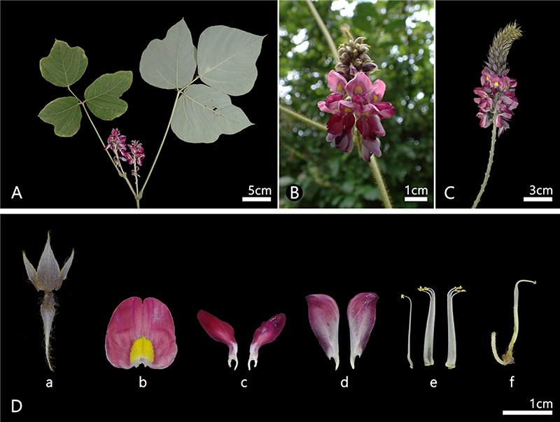
Photographs of P. lobata in natural populations. A; Plant habit, B-C; Inflorescence, D; Floral morphology, a; calyx, b; standard petal, c; wing petals, d; keel petals, e; stamen, f; pistil.
관찰 및 기재는 Ojeda et al. (2009)을 참조하여 화판을 기판 (standard), 익판 (wings), 용골판 (keels)으로 구분하고, 각 화판을 외면 (outer = abaxial side)과 내면 (inner = adaxial side)으로 나누어 관찰하였다. 또한 근위 (proximal), 원위 (distal), 축 (axis)을 따라 기저부 (base), 중앙부 (middle), 선단부 (apical)로, 중앙부 (medial)에서는 측면 (lateral) 축에 따라 중앙 (center)와 가장자리 (margin)로 구분하였다. 모든 시료는 주사전자현미경 (scanning electron microscope, EM30+, COXEM, Daejeon, Korea)을 이용하여 가속전압 5 kV, 작동거리 5.0—7.0 ㎜에서 관찰하였으며, 1,000—5,000배 배율로 촬영하였다.
결 과
1. 칡의 외부형태학적 형질
칡은 덩굴성 목본 (climbers, vine)이고, 잎은 3출 복엽 (trifoliolate)이며, 정소엽은 갈라지지 않거나, 3갈래로 갈라지는 것으로 관찰되었다 (Fig. 1A). 화서 (inflorescences)는 총상 (raceme)으로 액생 (axillary)하고, 화축 (rachis)에서는 돌출된 마디 (nodes)가 관찰되며, 포 (bracts)는 소형으로 조락성 (caducous)이고, 개화 후 빠르게 탈락하는 것으로 확인되었다 (Fig. 1A-C). 악 (calyx)은 종형 (campanulate)으로 전체적으로 황색 털이 산생하였으며, 열편 (lobes)은 5개이나, 상부의 두 열편이 동합 (connate)되어 4개로 보이고, 하부 열편은 상부 열편에 비해 길이가 약 두 배 긴 것으로 확인되었다 (Fig. 1Da). 화판 (petals)은 적자색으로 관찰되었으며, 기판 (standard)은 기부에는 뚜렷한 돌기가 존재하고, 중심부에는 황색 무늬가 관찰되었다. 익판 (wings)은 도란형 (obovate) 내지 장타원형 (oblong)으로 관찰되었으며, 용골판 (keels)은 장타원형 (oblong)으로 관찰되었고, 익판과 용골판의 길이는 유사한 것으로 확인되었다 (Fig. 1Db-d). 수술은 양체웅예 (diadelphous stamen, 9+1)이며, 약 (anthers)은 동형 (isomorphic)으로 관찰되었고 (Fig. 1De), 암술은 1개이며, 자방은 선형 (linear)으로 관찰되었다 (Fig. 1Df).
2. 기판의 미세형태
화판 표피세포 형태는 다음의 네 가지 유형으로 구분하였다 : 원추형 유두상 세포형 (PCS, Papillose conical cells with striations, Fig. 2A), 혹상 유두상 세포형 (PKR, Papillose knobby cells with a rugose sculpture, Fig. 2B), 주름판상 세포형 (TRS, Tabular rugose cells with longitudinal striations, Fig. 2C), 줄무늬 평판상 세포형 (TFS, Tabular flat cells with longitudinal striations, Fig. 2D) (Table 1; Fig. 2).
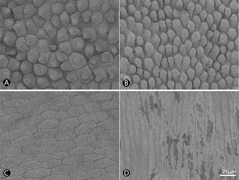
SEM micrographs of the cell types observed in P. lobata and Puerariae Flos. A; PCS, papillose conical cells with striations, B; PKR, papillose knobby cells with a rugose sculpture, C; TRS, tabular rugose cells with longitudinal striations, D; TFS, tabular flat cells with striations, A, C; P. lobata, B, D; Puerariae Flos.

Micromorphological characteristics of petals in P. lobata and Puerariae Flos.
칡과 갈화의 기판 외면을 비교한 결과, 칡의 선단부와 중앙부에서 원추형 유두상 세포 (PCS)가 관찰되었으며 (Fig. 3A, E), 기저부에서는 주름판상 세포 (TRS)가 확인되었다 (Fig. 3I). 반면, 갈화의 선단부와 중앙부에서 혹상 유두상 세포 (PKR)가 나타났으며 (Fig. 3C, G), 기저부에서는 주름판상 세포 (TRS)가 관찰되었다 (Fig. 3K). 또한 수층벽은 두 시료 모두에서 굽은형 (curved)으로 나타났고, 큐티클 표면은 유선(striate)이 공통적으로 확인되었다.
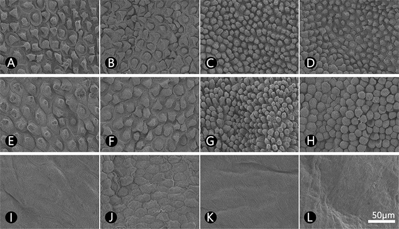
SEM micrographs of the central region of the standard petal surface in P. lobata and Puerariae Flos. A, C, E, G, I, K; Outer surface, B, D, F, H, J, L; Inner surface, A-D; Apical region, E-H; Middle region, I-L; Base region, A-B, E-F, I-J; P. lobata, C-D, G-H, K-L; Puerariae Flos.
내면에서는 칡의 선단부와 중앙부에서 원추형 유두상 세포 (PCS)가 확인되었고 (Fig. 3B, F), 기저부에서는 주름판상 세포 (TRS)가 관찰되었다 (Fig. 3J). 반면 갈화에서는 선단부와 중앙부에서 혹상 유두상 세포 (PKR)가 나타났으며 (Fig. 3D, H), 기저부에서는 줄무늬 평판상 세포 (TFS)가 확인되었다 (Fig. 3L). 수층벽은 칡에서 전반적으로 굽은형으로 나타났고, 갈화에서도 선단부와 중앙부에서 굽은형이 관찰되었으나, 기저부에서는 큐티클에 의해 덮여 수층벽이 명확히 확인되지 않았다. 큐티클 표면은 칡 전체와 갈화의 선단부 및 중앙부에서 유선이 확인되었고, 갈화의 기저부에서는 직선형이 확인되었다.
3. 익판의 미세형태
칡과 갈화의 익판 외면 세포형태를 비교한 결과, 칡에서는 선단부와 중앙부에서 원추형 유두상 세포 (PCS)가 관찰되었으며 (Fig. 4A, E), 기저부에서는 주름판상 세포 (TRS)가 확인되었다 (Fig. 4I). 반면 갈화에서는 전 영역에서 혹상 유두상 세포 (PKR)가 관찰되었다 (Fig. 4C, G, K). 수층벽은 두 시료 모두에서 굽은형으로 나타났다. 큐티클 표면의 경우, 칡은 선단부와 중앙부에서 유선형의 유선이 발견되었고, 기저부에서는 직선형의 유선이 관찰되었으며, 갈화에서는 전반적으로 유선형의 유선만 확인되었다. 익판 내면에서는 칡의 전 영역에서 원추형 유두상 세포 (PCS)가 관찰되었고 (Fig. 4B, F, J), 갈화에서는 혹상 유두상 세포 (PKR)가 확인되었다 (Fig. 4D, H, L). 수층벽은 두 시료 모두에서 굽은형으로 나타났으며, 큐티클 표면에서도 공통적으로 유선형의 유선이 나타났다.
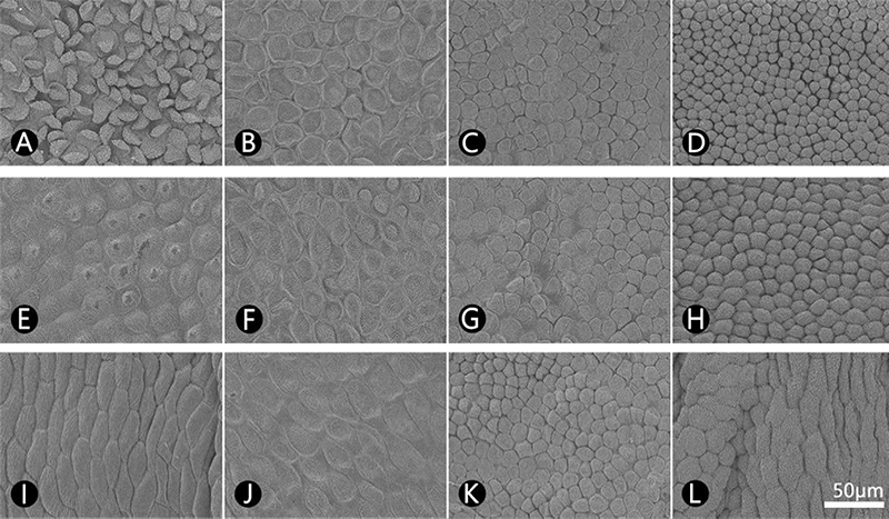
SEM micrographs of the central region of the wing petal surface in P. lobata and Puerariae Flos. A, C, E, G, I, K; Outer surface, B, D, F, H, J, L; Inner surface, A-D; Apical region, E-H; Middle region, I-L; Base region, A-B, E-F, I-J; P. lobata, C-D, G-H, K-L; Puerariae Flos.
4. 용골판의 미세형태
칡과 갈화의 용골판 외면 세포 형태를 비교한 결과, 칡에서는 선단부와 중앙부에서 주름판상 세포 (TRS)가 관찰되었으나 (Fig. 5A, E), 기저부에서는 세포 경계가 불명확하여 세포 형태, 수층벽과 큐티클이 뚜렷하게 확인되지 않았다 (Fig. 5I). 반면 갈화에서는 주름판상 세포 (TRS)가 일관되게 확인되었다 (Fig. 5C, G, K). 수층벽은 두 시료 모두에서 굽은형으로 나타났다. 큐티클 표면은 칡의 선단부와 중앙부에서 유선형으로 관찰되었으며, 갈화에서도 대부분 유선형으로 확인되었다. 다만, 칡에서는 선단부에서 중앙부로 갈수록 유선의 밀도가 감소하는 경향을 보인 반면, 갈화에서는 이러한 밀도 차이가 뚜렷하게 나타나지 않았다. 기저부에서는 칡의 경우 큐티클 표면이 확인되지 않았고, 갈화에서는 유선 밀도가 매우 낮게 관찰되었다.
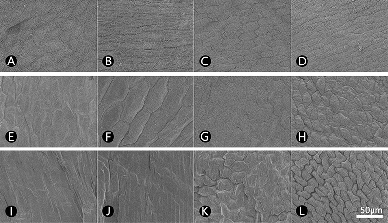
SEM micrographs of the central region of the keel petal surface in P. lobata and Puerariae Flos. A, C, E, G, I, K; Outer surface, B, D, F, H, J, L; Inner surface, A-D; Apical region, E-H; Middle region, I-L; Base region, A-B, E-F, I-J; P. lobata, C-D, G-H, K-L; Puerariae Flos.
내면에서는 칡은 선단부에 줄무늬 평판상 세포 (TFS), 중앙부에서 주름판상 세포 (TRS)가 관찰되었으나 (Fig. 5B, F), 기저부에서는 세포 간 경계가 불명확하여 세포 형태, 수층벽과 큐티클의 확인이 어려웠다 (Fig. 5J). 반면 갈화에서는 내면 전 영역에서 주름판상 세포 (TRS)가 일관되게 관찰되었다 (Fig. 5D, H, L). 수층벽은 칡의 선단부와 중앙부에서는 굽은형으로 관찰되었으며, 갈화 역시 선단부와 중앙부에서는 굽은형을 나타냈으나 기저부에서는 파상형 (sinuous)으로 관찰되었다. 큐티클 표면의 경우, 칡의 선단부와 중앙부에서는 직선형과 유선이 함께 확인되었고, 갈화는 선단부와 중앙부에서 유선형의 유선이 관찰되었으며, 기저부에서는 직선형의 유선이 관찰되었다.
고 찰
본 연구에서는 채집 시료인 칡과 유통 약재 시료인 갈화의 화판 양면에서 표피세포, 수층벽 및 큐티클의 미세형태학적 형질을 주사전자현미경을 이용하여 자세히 기재하고, 비교 및 검토하였다.
화판의 미세형태학적 분석 결과, 칡과 갈화의 기판과 익판 모두에서 유두상 세포 (papillose cells)가 관찰되었다. 유두상 세포는 꽃과 수분 매개자 간의 상호작용에 중요한 역할을 하는 구조로 알려져 있다 (Kay et al., 1981; Christensen and Hansen, 1998). 특히 벌과 같은 수분 매개자는 유두상 세포가 발달한 꽃을 선호하는 경향이 있으며, 이는 유두상 세포가 화판 표면의 부착력을 증가시켜 안정적인 착지 기반을 제공함으로써 먹이 활동 시 에너지 소모를 줄여주기 때문으로 해석된다 (Whitney et al., 2009; Alcorn et al., 2012). 또한 유두상 세포는 빛을 색소체로 집중시키는 렌즈와 같은 기능을 수행하여 꽃의 색을 더욱 선명하고 강렬하게 만들어 수분 매개자를 유인하는 시각적 신호로 작용한다고 보고되어 있다 (Noda et al., 1994; Gorton and Vogelmann, 1996; Glover and Martin, 1998; Comba et al., 2000).
콩과 식물에서는 일반적으로 수분 매개자가 인지하는 위치에 해당하는 기판과 익판에 유두상 세포가, 상대적으로 인지 필요성이 낮은 용골판에는 판상 세포 (tabular cells)가 분포하는 것으로 보고되어 있으며 (Ojeda et al., 2009; Ojeda et al., 2012), 이러한 세포 유형의 조합과 분포 패턴은 종 및 아종 수준에서 비교적 일관된 진단 형질로 활용될 수 있다 (Çildir et al., 2012). 본 연구에서도 칡과 갈화의 표피세포 형태는, 기판과 익판에서는 유두상 세포가, 용골판에서는 판상 세포가 관찰되어 기존 연구 결과를 지지하였다.
채집 시료와 약재 시료에서 표피세포의 형태는 전반적으로 유사하였으나, 유두상 세포의 형태에서 시료 간 차이가 확인되었다. 기존 감별 연구에서는 화분·포자 약재의 표면 미세형태 (Doh et al., 2015; Ramzan et al., 2019; Kim et al., 2020; Bahadur et al., 2022), 잎 표피 미세·내부형태 (Song et al., 2020; Stoyanov et al., 2025), 뿌리 내부형태 (Ji et al., 2026) 등 다양한 기관의 외부·내부·미세형태 형질은 진품과 위품 (adulterant)을 판별하는 유용한 지표로 활용되어 왔다. 특히 콩과 식물에서는 유두상 세포의 형태, 표면 무늬 (cuticular relief), 그리고 세포 배열 양상이 비교적 안정적으로 유지되는 것으로 알려져 있다. 이는 이러한 형질이 발달과 밀접하게 연관된 구조적 특성이기 때문에 환경 요인에 의한 변이가 제한되기 때문으로 해석된다 (Ojeda et al., 2009). 따라서 본 연구에서 확인된 유두상 세포 형태의 차이는 채집 시료와 약재 시료 간에 존재하는 미세형태학적 변이를 반영하는 것으로 해석될 수 있다. 이러한 변이는 수분 매개자와의 상호작용, 환경 조건, 생장 단계, 또는 분류학적 차이 등 다양한 요인과 관련될 가능성이 있으나, 현재 자료만으로 그 원인을 명확히 구분하기는 어렵다. 특히 본 연구에 사용된 약재 시료는 중국산으로, 칡 (P. lobata) 한 종만이 분포하는 한국과 달리 중국에는 약 10종의 동속 근연종이 분포하는 것으로 알려져 있어 (Wu and Thulin, 2010), 해당 시료가 칡 이외의 동속 이종일 가능성도 배제할 수 없다. 그러나 현재까지 본 식물에서 유두상 세포 형태와 수분 매개자 간의 상호작용을 직접적으로 규명한 연구는 보고된 바가 없으며, 시료 수 또한 제한적이기 때문에 관찰된 형태적 차이가 환경적 요인이나 생장 단계에 따른 변이인지, 혹은 종간 분류학적 차이에 기인한 것인지를 단정하기에는 한계가 있다. 따라서 이러한 미세형태학적 차이의 기원을 명확히 규명하기 위해서는 보다 다양한 분류군과 시료를 포함하고, 생태적·발생학적 요인을 통합적으로 고려한 후속 연구가 필요하다.
단일 화판 내에 두 가지 이상의 주요 표피 유형이 공존하는 현상은 일부 제한된 분류군에서 보고된 바 있다 (Baagøe, 1977; Barthlott and Ehler, 1977; Hansen, 1991; Christensen and Hansen, 1998). 특히 콩아과 (Faboideae Rudd)의 일부 분류군에서는 하나의 화판 표면에 서로 뚜렷이 구별되는 두 가지 표피 유형이 동시에 나타나며, 이들 사이에는 전이대 (transition zone)라는 중간 형태의 세포 영역이 존재하는 것으로 보고되었다 (Ojeda et al., 2009). 본 연구에서도 채집 시료의 익판 외면에서 선단부에서 기저부 방향으로 갈수록 원추형 유두상 세포형 (PCS)이 주름판상 세포형 (TRS)으로 점진적으로 변화하는 양상이 관찰되었으며, 이 과정에서 중앙부에는 전이대에 해당하는 중간형 세포 영역이 확인되었다 (Fig. 4E). 반면, 약재 시료에서는 혹상 유두상 세포형 (PKR)만이 관찰되었고, 전이대는 확인되지 않았다. 이러한 구조적 차이는 시료 간 미세형태학적 변이를 반영하는 것으로 해석될 수 있으나, 그 원인이 분류학적 차이에 기인하는지 여부는 현재 자료만으로는 명확히 판단하기 어렵다. 따라서 이러한 형질의 분포 양상과 그 기원을 이해하기 위해서는 보다 다양한 분류군과 충분한 시료를 포함한 추가적인 검증 연구가 필요하다.
화판의 미세형태는 단일 화판 내에서도 근위–원위부 축을 따라 분화 정도에 차이가 있는 것으로 알려져 있다. 일반적으로 화판의 기저부는 분화가 덜 된 세포로 구성되어 종 고유의 세포 형태가 뚜렷하게 나타나지 않는 반면, 선단부와 중앙부에서는 분화가 완료된 세포가 관찰되어 종 특이적 형태가 보다 명확히 드러나는 것으로 보고되어 왔다 (Ojeda et al., 2009). 그러나 본 연구에서 채집 시료와 약재 시료의 화판을 미세형태학적으로 분석한 결과, 이러한 일반적 경향과는 다소 상이하게 기저부 영역에서도 분화가 진행된 것으로 보이는 세포 형태가 관찰되었다 (Figs. 3I-L, 4I-L, 5I-L). 다만, 이러한 기저부 세포의 형태는 모든 시료에서 일관되게 나타나지 않았으며, 특히 칡의 용골판 기저부에서는 세포 경계가 불명확한 비교적 단순한 형태가 관찰되었다 (Fig. 5I-J). 이러한 변이가 종 고유의 형질을 반영하는지, 또는 환경적 요인이나 약재 가공 과정에 의해 유발된 것인지는 현재 자료만으로는 명확히 판단하기 어렵다. 따라서 이러한 관찰 결과를 일반화하기 위해서는 칡속의 다양한 분류군과 충분한 시료를 포함한 추가적인 검증 연구가 필요하다. 나아가 향후 칡속 전반을 대상으로 한 화판 표피세포의 미세형태학적 비교가 이루어진다면, 이러한 형질의 변이 양상에 대한 보다 정밀한 이해가 가능할 것으로 기대된다.
화판 표피세포는 국화과 (Batista and De Souza, 2017; Gavrilović and Janaćković, 2022), 장미과 (Song and Hong, 2016; Seyedipour and Mahdigholi, 2024) 등 다양한 분류군의 계통분류학적 연구에 활용되어 왔으며, 콩아과 내에서도 화판 표피의 미세형태는 분류학적 유용성을 지니는 형질로 보고된 바 있다 (Ojeda et al., 2009). 본 연구에서는 칡의 화판에서 원추형 유두상 세포 (PCS)가 우세하게 관찰된 반면, 갈화에서는 혹상 유두상 세포 (PKR)가 주된 세포 형태로 나타났다. 이러한 미세형태학적 차이는 시료 간 변이를 반영하는 것으로 해석될 수 있으며, 칡속 내 근연 분류군의 구분이나 칡과 갈화의 감별에 활용될 가능성을 시사한다. 다만, 본 연구 결과는 제한된 시료에 기반한 것이므로, 이러한 형질의 일반성과 재현성을 평가하기 위해서는 추가적인 검증이 필요하다. 또한 본 연구에서 제시한 미세형태학적 형질의 일관성을 확인하기 위한 유통 약재 모니터링 역시 요구된다. 한편, 유통 약재의 정확한 종 동정을 위해 DNA 바코딩 등 분자계통학적 분석과 이화학적 지표를 포함한 다양한 정보를 통합적으로 활용한 후속 연구가 필요하다. 이러한 접근은 갈화와 칡의 감별 정확도 향상에 기여할 수 있을 뿐만 아니라, 콩아과 내 미세형태학적 형질의 분류학적 활용 가능성을 보다 정밀하게 평가하는 데에도 도움이 될 것으로 기대된다.
본 연구에서는 채집 시료인 칡과 유통 약재인 갈화를 대상으로 화판 표피의 미세형태를 비교·분석하였으며, 특히 콩과 식물의 화판을 기판, 익판, 용골판 및 위치별로 구분하여 정밀하게 관찰하였다는 점에서 의의가 있다. 두 시료 모두에서 관찰된 표피세포 유형은 콩아과에서 보고된 일반적인 경향과 전반적으로 일치하였다. 다만, 채집 시료에서는 원추형 유두상 세포 (PCS), 약재 시료에서는 혹상 유두상 세포 (PKR)가 상대적으로 우세하게 나타나 시료 간 미세형태학적 차이가 확인되었다. 이러한 차이는 화판 표면의 기능적 특성과 관련될 가능성을 시사하지만, 그 생물학적 의미나 원인에 대해서는 현재 자료만으로 명확히 해석하기 어렵다.
또한 칡의 익판에서는 원추형 유두상에서 주름판상으로의 변화와 전이대가 관찰된 반면, 약재 시료에서는 이러한 전이대가 확인되지 않았다. 이러한 관찰 결과는 시료 간 형태적 변이를 보여주지만, 그 원인이 분류학적 차이에 기인하는지 여부는 단정하기 어렵다. 특히 유통 약재의 경우 다양한 기원종 및 가공 과정의 영향을 받을 가능성이 있으므로 보다 폭넓은 시료를 대상으로 한 추가적인 검증이 필요하다.
따라서 본 연구에서 제시한 미세형태학적 특징들은 갈화와 칡의 구분을 위한 잠재적 형질로서 활용 가능성을 제시하는 기초자료로 의미가 있으며, 향후 다양한 분류군과 충분한 시료를 포함한 반복 연구와 함께 분자계통학적 및 이화학적 분석을 병행한 통합적 접근이 요구된다. 이러한 후속 연구는 갈화의 기원종 규명과 약재의 품질 관리 기준 설정에 기초 자료를 제공할 수 있을 것으로 기대된다.
Acknowledgments
본 연구는 한국연구재단 G-램프 (LAMP) 사업 (Grant No. RS-2024-00445180) 및 산림청 (한국임업진흥원) 산림과학기술 연구개발사업 (RS-2021-KF001796)의 지원을 받아 수행되었으며, 이에 감사드립니다.
References
-
Alcorn K, Whitney H and Glover B. (2012). Flower movement increases pollinator preference for flowers with better grip. Functional Ecology. 26:941-947.
[https://doi.org/10.1111/j.1365-2435.2012.02009.x]

- Aswini A, Kumarasamy D, Lazar J, Deebika K, Arun VP and Nahendran C. (2024). Exploring pollen morphology diversity within the Fabaceae family: A study from Annamalai Nagar, Chidambaram. International Journal of Biosciences. 25:93-105.
- Baagøe J. (1977). Taxonomic application of ligule microcharacters in Compositae. Svensk Botanisk Tidskrift. 71:193-223.
-
Bahadur S, Taj S, Ahmad M, Zafar M, Gul S, Shuaib M, Shah SN, Begum N, Ramamneh EA and Romman M. (2022). Authentication of the therapeutic Lamiaceae taxa by using pollen traits observed under scanning electron microscopy. Microscopy Research and Technique. 85:2026-2044.
[https://doi.org/10.1002/jemt.24061]
- Barthlott W. (1984). Microstructural features of seed surfaces. In Heywood VH and Moore DM. (ed.). Current concepts in plant taxonomy. Academic Press. London, UK. p.95-105.
- Barthlott W and Ehler N. (1977). Raster-Elektronenmikroskopie der Epidermis-Oberflächen von Spermatophyten(Tropische und Subtropische Pflanzenwelt, Vol. 19). Franz Steiner Verlag. Wiesbaden, Germany. p.367-467.
-
Batista MF and De Souza LA. (2017). Flower structure in ten Asteraceae species: Considerations about the importance of morpho-anatomical features at species and tribal level. Brazilian Journal of Botany. 40:265-279.
[https://doi.org/10.1007/s40415-016-0312-9]
- Choi BH. (2018). Fabaceae. In Flora of Korea Editorial Committee (eds.). The Genera of Vascular Plants of Korea. Hongreung Publishing Co. Seoul, Republic of Korea. p.770-820.
- Choi BH. (2024). Fabaceae Lindl.(Leguminosae Juss.) In Flora of Korea Editorial Committee (eds.). Flora of Korea. Volume 5a-3(Rosidae: Fabaceae). Junghaengsa. Incheon, Republic of Korea. p.3-87.
- Christensen KI and Hansen HV. (1998). SEM studies of epidermal patterns of petals in angiosperms. Opera Botanica. 135:5-91.
-
Comba L, Corbet SA, Hunt H, Outram S, Parker JS and Glover BJ. (2000). The role of genes influencing the corolla in pollination of Antirrhinum majus. Plant, Cell & Environment. 23:639-647.
[https://doi.org/10.1046/j.1365-3040.2000.00580.x]
-
Çildir H, Kahraman A and Dogan M. (2012). Petal and sepal epidermal micromorphology of six Lathyrus taxa(Fabaceae) and their systematic value. Notulae Botanicae Horti Agrobotanici Cluj Napoca. 40:87-93.
[https://doi.org/10.15835/nbha4017503]
-
Doh EJ, Kim JH, Choi G, Lee SH, Song HJ, Ju YS and Lee GS. (2015). Microscopic identification-keys for Cynanchi Wilfordii Radix and Cynanchi Auriculati Radix. The Korea Journal of Herbology. 30:65-69.
[https://doi.org/10.6116/kjh.2015.30.4.65.]
-
Eggli U and Clark RP. (2023). Fabaceae. In Eggli U and Nyffeler R. (eds.). Dicotyledons: Rosids. Illustrated Handbook of Succulent Plants. Springer. Cham, Switzerland. p.645-653.
[https://doi.org/10.1007/978-3-030-93492-7_61]
-
Gavrilović M and Janaćković P. (2022). Micromorphology of endemic Centaurea glaberrima subsp. divergens(Asteraceae). Acta Botanica Croatica. 81:23-31.
[https://doi.org/10.37427/botcro-2021-028]
-
Glover BJ and Martin C. (1998). The role of petal cell shape and pigmentation in pollination success in Antirrhinum majus. Heredity. 80:778-784.
[https://doi.org/10.1046/j.1365-2540.1998.00345.x]
-
Gorton HL and Vogelmann TC. (1996). Effects of epidermal cell shape and pigmentation on optical properties of Antirrhinum petals at visible and ultraviolet wavelengths. Plant Physiology. 112:879-888.
[https://doi.org/10.1104/pp.112.3.879]
-
Hai Y, Huang X, Sun H, Sun J, Li J, Zhang Y, Li Y, Wu P and Xia C. (2024). Comparative analysis of the complete chloroplast genome of Pueraria provides insights for species identification, phylogenetic relationships, and taxonomy. BMC Plant Biology. 24:1196. (cited by 2026 May 18).
[https://doi.org/10.1186/s12870-024-05905-9]
- Hansen HV. (1991). Phylogenetic studies in Compositae tribe Mutisieae. Opera Botanica. 109:1-50.
-
He H, Peng S, Song X, Jia R, Zou Y, Li L and Yin Z. (2022). Protective effect of isoflavones and triterpenoid saponins from Pueraria lobata on liver diseases: A review. Food Science & Nutrition. 10:272-285.
[https://doi.org/10.1002/fsn3.2668]
- IPNI. (2026). International Plant Names Index. The Royal Botanic Gardens, Kew, Harvard University Herbaria & Libraries and Australian National Herbarium. http://www.ipni.org, . (cited by 2026 May 18).
-
Ji H, Choi WR, Choi JW and Song J-H. (2026). Comparative study on the morphological and anatomical characteristics of Trichosanthes kirilowii var. kirilowii and T. kirilowii var. japonica(Cucurbitaceae). Korean Journal of Medicinal Crop Science. 34:105-116.
[https://doi.org/10.7783/KJMCS.2026.34.2.105]
-
Kay QON, Daoud HS and Stirton CH. (1981). Pigment distribution, light reflection and cell structure in petals. Botanical Journal of the Linnean Society. 83:57-84.
[https://doi.org/10.1111/j.1095-8339.1981.tb00129.x]
- Kim YS, Song J-H, Choi G, Lee G and Ju YS. (2020). Comparative study on the external micro-morphology of 3 kinds of minute pollen and spore Herbs(Pini Pollen, Typhae Pollen, Lygodii Spora) utilizing scanning electron microscope. The Korea Journal of Herbology. 35:9-18.
-
Lee HY, Jeon YJ, Kong MJ and Song J-H. (2026). A comparative study of floral and pollen micromorphology in Robinia pseudoacacia and Robiniae Flos(Fabaceae). Korean Journal of Medicinal Crop Science. 34:151-160.
[https://doi.org/10.7783/KJMCS.2026.34.3.151]
-
Lee JM, Kang YC and Song J-H. (2025). Comparative study of floral micromorphology from Rosae Rugosae Flos and Rosae Chinensis Flos(Rosaceae). Korean Journal of Medicinal Crop Science. 33:298-307.
[https://doi.org/10.7783/KJMCS.2025.33.5.298]
- Legume Phylogeny Working Group, LPWG (2017). A new subfamily classification of the Leguminosae based on a taxonomically comprehensive phylogeny. Taxon. 66:44-77.
- Lewis G, Schrire B, Mackinder B and Lock M. (2005). Legumes of the World. Royal Botanic Gardens. Kew. Richmond. UK. p.1-577.
- Ministry of Food and Drug Safety(MFDS). (2024). The Korean Herbal Pharmacopoeia(MFDS Notification No. 2024-85). Ministry of Food and Drug Safety. Cheongju, Republic of Korea.
-
Noda K, Glover BJ, Linstead P and Martin C. (1994). Flower colour intensity depends on specialized cell shape controlled by a MYB-related transcription factor. Nature. 369:661-664.
[https://doi.org/10.1038/369661a0]
- Ohwi J. (1947). Flora of Japan and its miscellaneous notes. Bulletin of the Tokyo Science Museum. 18:1-16.
-
Ojeda I, Francisco Ortega J and Cronk QC. (2009). Evolution of petal epidermal micromorphology in Leguminosae and its use as a marker of petal identity. Annals of Botany. 104:1099-1110.
[https://doi.org/10.1093/aob/mcp211]
-
Ojeda I, Santos-Guerra A, Caujapé-Castells J, Jaén-Molina R, Marrero A and Cronk QC. (2012). Comparative micro-morphology of petals in Macaronesian Lotus(Leguminosae) reveals a loss of papillose conical cells during the evolution of bird pollination. International Journal of Plant Sciences. 173:365-374.
[https://doi.org/10.1086/664713]
- Pan B, Yao X, Corlett RT and Yu WB. (2023). Pueraria omeiensis(Fabaceae), a new species from Southwest China. Taiwania. 68:31-38.
- Park JS, Kim KY and Han G. (2006). Ethanol toxicant composed of Pueraria thunbergiana and Alpinia katsumadai. Journal of the Pharmaceutical Society of Korea. 50:308-312.
-
Parveen I, Gafner S, Techen N, Murch SJ and Khan IA. (2016). DNA barcoding for the identification of botanicals in herbal medicine and dietary supplements: strengths and limitations. Planta Medica. 82:1225-1235.
[https://doi.org/10.1055/s-0042-111208]
- Polhill RM and Raven PH. (1981). Advances in legume systematics(Parts 1 and 2). Royal Botanic Gardens. Kew, UK. p.1-1050.
- POWO. (2026). Plants of the World Online. Royal Botanic Gardens, Kew. https://powo.science.kew.org/, (cited by 2026 May 18).
-
Ramzan S, Shaheen S, Hussain K, Sharifi-Rad J, Khan MA, Harun N, Zafar M and Khan F. (2019). Implication of palynological techniques for the authentication of adulterated drugs traded with the same name in different herbal markets of district Lahore, Pakistan. Microscopy Research and Technique. 82:931-940.
[https://doi.org/10.1002/jemt.23240]
- Sanjappa M. (1992). Legumes of India. Bishen Singh Mahendra Pal Singh. Dehra Dun, India. p.288.
-
Seyedipour S and Mahdigholi K. (2024). Petal micromorphology of some Rosa species in Iran. Rostaniha. 25:123-138.
[https://doi.org/10.1016/j.bse.2025.105056]
-
Song J-H and Hong SP. (2016). A study on the petal micromorphological characteristics of the tribe Sorbarieae (Rosaceae). Korean Journal of Plant Resources. 29:376-384.
[https://doi.org/10.7732/kjpr.2016.29.4.376]
-
Song J-H, Yang S and Choi G. (2020). Taxonomic implications of leaf micromorphology using microscopic analysis: A tool for identification and authentication of Korean Piperales. Plants. 9:566. (cited by 2026 May 18).
[https://doi.org/10.3390/plants9050566]
- Stoyanov AN, Gruykin TM, Boyadzhiev MY and Popovski VV. (2025). Pharmacognostic automated identification of folia. Comptes rendus de l'Académie bulgare des Sciences. 73:1555-1564.
- van der Maesen LJG. (1985). Revision of the genus Pueraria DC. with some notes on Teyleria Backer. Agricultural University Wageningen Papers. 85:1-132.
-
van der Valk JM, Leon CJ and Nesbitt M. (2017). Macroscopic authentication of Chinese materia medica(CMM): a UK market study of seeds and fruits. Journal of Herbal Medicine. 8:40-51.
[https://doi.org/10.1016/j.hermed.2017.03.007]
-
Wang H, Zou H, Cheng C, Wang W, Wang S, Wang D and Liu X. (2023). Authentication of Bistortae Rhizoma and its three adulterants based on their macroscopic morphology and microscopic characteristics. Microscopy Research and Technique. 86:359-367.
[https://doi.org/10.1002/jemt.24277]
-
Wang Z, Du H, Peng W, Yang S, Feng Y, Ouyang H, Xu S and Liu R. (2022). Efficacy and mechanism of Pueraria lobata and Pueraria thomsonii polysaccharides in the treatment of type 2 diabetes. Nutrients. 14:3926. (cited by 2026 May 18).
[https://doi.org/10.3390/nu14193926]
-
Whitney HM, Chittka L, Bruce TJA and Glover BJ. (2009). Conical epidermal cells allow bees to grip flowers and increase foraging efficiency. Current Biology. 19:948-953.
[https://doi.org/10.1016/j.cub.2009.04.051]
- Wilkinson HP. (1979). The plant surface(mainly leaf). In Metcalfe CR and Chalk L. (eds.). Anatomy of the dicotyledons(Vol. 1). 2nd ed. Clarendon Press. Oxford, UK. p.97-165.
- Willdenow CL. (1802). Species plantarum(Vol. 3, Part 2). G. C. Nauk. Berlin, Germany. p.1047.
- Wu DL and Thulin M. (2010). Pueraria DC. In Wu ZY, Raven PH and Hong DY. (eds.). Flora of China(Vol. 10). Science Press and Missouri Botanical Garden Press. Beijing, China and St. Louis, Missouri, USA. p.244-248.
-
Zhao Z, Liang Z and Ping G. (2011). Macroscopic identification of Chinese medicinal materials: traditional experiences and modern understanding. Journal of Ethnopharmacology. 134:556-564.
[https://doi.org/10.1016/j.jep.2011.01.018]
-
Zhou Y, Shang XH, Xiao L, Wu ZD, Cao S and Yan HB. (2023). Comparative plastomes of Pueraria montana var. lobata(Leguminosae: Phaseoleae) and closely related taxa: Insights into phylogenomic implications and evolutionary divergence. BMC Genomics. 24:299. (cited by 2026 May 18).
[https://doi.org/10.1186/s12864-023-09356-8]