
Anti-inflammatory Effects and Gastric Mucosal Protection of Phyllostachyos Caulis in Taeniam Extract against EtOH/HCl-Induced Gastric Mucosal Injury
This is an open access article distributed under the terms of the Creative Commons Attribution Non-Commercial License (http://creativecommons.org/licenses/by-nc/3.0/) which permits unrestricted non-commercial use, distribution, and reproduction in any medium, provided the original work is properly cited.
Abstract
Phyllostachyos Caulis in Taeniam (PCT), commonly known as bamboo shavings, is a traditional medicinal herb widely used in Korea and China for its heat-clearing, phlegm-resolving, and anti-inflammatory properties. This study investigated the antioxidant, anti-inflammatory, and gastroprotective effects of the hot-water extract of PCT (PCTD).
PCTD exhibited radical-scavenging activity in DPPH and ABTS assays and anti-inflammatory effects in lipopolysaccharide (LPS)-stimulated RAW264.7 cells. Its gastroprotective effects were evaluated in a mouse model of acute gastric mucosal injury induced by EtOH/HCl. In LPS-stimulated RAW264.7 cells, PCTD reduced nitric oxide and prostaglandin E2levels without compromising cell viability. At a concentration of 200 ㎍/mL, PCTD also modulated the expression of inflammation-related cytokines and proteins. Furthermore, oral administration of PCTD significantly reduced gross mucosal injury in the EtOH/HCl-induced model. Representative histological sections revealed differences in mucosal morphology and PAS-positive and TUNEL-positive staining following PCTD treatment.
These findings indicate that PCTD exhibits antioxidant and anti-inflammatory activities in vitro and attenuates EtOH/HCl-induced acute gastric mucosal injury in mice. Its ability to suppress pro-inflammatory mediators in macrophages and protect against gastric mucosal injury highlights its therapeutic potential as a natural anti-inflammatory and gastroprotective agent.
Keywords:
Anti-inflammatory, Antioxidant, Gastritis, Phyllostachyos Caulis in Taeniam, Plant Resource, Traditional MedicineINTRODUCTION
Phyllostachyos Caulis in Taeniam (PCT) is derived from the middle layer of the stems of Phyllostachys nigra var. henonis or Phyllostachys bambusoides. PCT is a commonly used medicinal material in traditional Chinese and Korean medicine (Kim et al., 2013; An et al., 2025). Since ancient times, PCT has been utilized for clearing heat, resolving phlegm, relieving adverse gastric symptoms, stopping vomiting, and calming the liver and nerves (Kim et al., 2013; An et al., 2025). Modern pharmacological studies indicate that PCT contains multiple active compounds, such as chlorogenic acid, ferulic acid, p-coumaric acid, and triterpenoids, which contribute to its notable antioxidant and anti-inflammatory activities (Lim et al., 2017; An et al., 2025). Jin et al. (2012) indicated that the ethyl acetate fraction of bamboo significantly suppressed levels of inflammatory mediators such as TNF-α, IL-1β, and IL-6 in RAW264.7 macrophages stimulated by Porphyromonas gingivalis LPS. Furthermore, the fraction activated the Nrf2/p38 MAPK pathway and induced heme oxygenase-1 (HO-1) expression, suggesting a role in the regulation of oxidative stress and inflammatory responses (Jin et al., 2012). Its triterpenoid-enriched extract (EBS) has been reported to possess antitumor and anti-lipid peroxidation properties (Lu et al., 2010). These findings suggest that bamboo shavings may have potential as a natural source of antioxidant and anti-inflammatory agents.
Gastritis is a common gastrointestinal disorder characterized by inflammation and structural damage to the gastric mucosa and may occur in acute or chronic forms (Kim et al., 2023). Major causes include Helicobacter pylori, chronic heavy drinking, and long-term use of nonsteroidal anti-inflammatory drugs (NSAIDs) (Yang et al., 2024). Alcohol consumption not only stimulates and damages gastric epithelial cells, but excessive intake also weakens the mucosal barrier, making it more vulnerable to irritation (Oh et al., 2005; Kim et al., 2021). The gastric mucosa is protected by several defense mechanisms, including the mucus-bicarbonate barrier, prostaglandin synthesis, mucosal blood flow, antioxidant defense, and epithelial repair (Hagen, 2021). Impairment of these protective mechanisms can further aggravate mucosal erosion and inflammatory damage. Natural plant extracts have been found to maintain gastric mucosal integrity by modulating oxidative stress and inflammatory pathways (Lee et al., 2022; Chen et al., 2025). This study aimed to confirm the anti-inflammatory activity of PCTD at the cellular level and investigate its inhibitory effects on inflammation and its protective effects on the gastric mucosa using an EtOH/HCl-induced acute gastric mucosal injury model.
MATERIALS AND METHODS
1. Plant material and extract
Phyllostachyos Caulis in Taeniam (PCT) used in this experiment was purchased from Kwangmyungdang Pharmaceutical Co. (Ulsan, South Korea), a product certified under the Good Manufacturing Practice (GMP) standards of the Ministry of Food and Drug Safety, South Korea. Documentation of the plant material includes a voucher specimen (No. 2-20-0025) deposited in the Korean Herbarium of Standard Resources at the Korea Institute of Oriental Medicine (KIOM) (Supplementary Fig. S1). The PCT was subjected to reflux extraction in distilled water for 3 hours. The solution was concentrated under reduced pressure, and freeze-dried. The extract was stored at –70℃.
2. Antioxidant activity
To evaluate the antioxidant activity of PCTD, the DPPH (2,2-diphenyl-1-picrylhydrazyl) radical scavenging assay was performed. PCTD was prepared at working concentrations of 25, 50, and 100 ㎍/mL. Gallic acid was used as a positive control at the same concentrations (25, 50, and 100 ㎍/mL). A volume of 100 μL of 0.2 mM DPPH solution was mixed with an equal volume of PCTD solution or gallic acid solution in a 96-well plate. The mixture was incubated in the dark for 30 minutes (Nam et al., 2021). Absorbance was measured at 517 ㎚ using a microplate spectrophotometer (SpectraMax i3x, Molecular Devices, San Jose, CA, USA).
To evaluate the antioxidant capacity of PCTD, the ABTS [2,2'-azinobis-(3-ethylbenzothiazoline-6-sulfonic acid)] radical scavenging assay was employed (Bibi Sadeer et al., 2020). A 7 mM ABTS solution was mixed with an equal volume of 2.45 mM potassium persulfate solution and reacted in the dark. The mixture’s absorbance at 734 ㎚ was quantified using a microplate reader, proceeding with the assay only when readings reached 0.70 ± 0.02. Subsequently, 180 μL of ABTS+ working solution was mixed with PCTD at various concentrations (25, 50, 100 ㎍/mL) and reacted for 10 min. Gallic acid was used as a positive control at the same concentrations (25, 50, and 100 ㎍/mL). After the reaction, absorbance was measured at 734 ㎚ using a microplate spectrophotometer (SpectraMax i3x, Molecular Devices, San Jose, CA, USA).
3. Anti-inflammatory activity
RAW264.7 macrophages, obtained from the American Type Culture Collection (ATCC, Manassas, VA, USA), were maintained in Dulbecco's Modified Eagle Medium (DMEM) containing 10% fetal bovine serum (FBS) along with 1% penicillin-streptomycin (P/S). Cells were cultured at 37℃ with 5% CO2 and used during the exponential growth phase.
RAW 264.7 cells were seeded in 96-well plates at a density of 2 × 10⁵ cells/well and incubated for 24 h. The cells were pretreated with PCTD (50, 100, or 200 ㎍/mL) for 1 h and then stimulated with 1 ㎍/mL lipopolysaccharide (LPS) for 24 h. Cell viability was assessed using the MTS assay by measuring absorbance at 490 ㎚. For NO analysis, nitrite levels in the culture supernatant were measured using Griess reagent at 540 ㎚ and calculated from a nitrite standard curve.
RAW264.7 cells were pretreated with PCTD (100 or 200 μg/mL) and then stimulated with LPS (1 μg/mL). After 24 h, supernatants were collected for analysis. To assess the effects of LPS and PCTD on PGE2 production, an ELISA kit (R&D Systems, Minneapolis, MN, USA) was used. Absorbance was measured at 450 ㎚ using a SpectraMax i3x microplate spectrophotometer (Molecular Devices, San Jose, CA, USA), and PGE2 production was calculated based on the standard curve.
The concentrations of IL-1β, IL-6, G-CSF, and GM-CSF in the cell culture supernatants were quantified using specific commercial ELISA kits (R&D Systems, Minneapolis, MN, USA) following the manufacturer's protocols. Cytokine concentrations are expressed as pg/mL.
RAW264.7 cells were pretreated with PCTD (100 or 200 μg/mL) for 1 h and then stimulated with LPS (1 μg/mL) for 18 h. Total cellular proteins were extracted, and the expression levels of COX-2, COX-1, IL-1β, and IL-10 were analyzed by western blotting. β-actin was used as a loading control. Protein band intensities were quantified using Image Lab software (Bio-Rad Laboratories, Hercules, CA, USA) and normalized to β-actin. The experiment was independently performed three times.
4. Protective effect against gastric mucosal injury
Six-week-old C57BL/6 male mice (20 ± 2 g) were purchased from Doolyul Bio (Seocho-gu, Seoul, Korea). After one week of acclimatization under standard laboratory conditions (temperature 22 ± 2℃, humidity 50 ± 5%, 12-hour light/dark cycle) with free access to food and water, they were used for experiments. All experimental equipment underwent sterilization. All animal experiments complied with global welfare guidelines and received formal approval from the KIOM Animal experiment ethics committee under protocol number (Approval No: 22-080).
To evaluate the gastroprotective effect of PCTD, gastric mucosal injury was induced by administration of 70% ethanol containing 150 mM HCl. To analyze changes in gastric mucosal injury severity, 24 mice were randomly divided into four groups (n = 6 per group): Normal control group (NC, receiving saline), EtOH/HCl group (receiving EtOH/HCl), positive control group (R25, receiving EtOH/HCl + ranitidine 25 ㎎/㎏), and PCTD-treated group (PCTD50, receiving EtOH/HCl + PCTD 50 ㎎/㎏). For one week before induction, the NC and EtOH/HCl groups received saline, the R25 group received ranitidine (25 ㎎/㎏), and the PCTD50 group received PCTD (50 ㎎/㎏) orally. The 50 ㎎/㎏ dose used in the present study falls within the range previously evaluated for Bambusae Caulis in Taeniam extracts in vivo (Ra et al., 2010; Lim et al., 2017). This pretreatment protocol was designed to evaluate the protective effect of PCTD against acute gastric mucosal injury rather than the post-injury recovery process. On the day of the experiment, after a 4-hour fast, the respective pretreatments (saline, ranitidine, PCTD) were administered. After two hours, EtOH/HCl solution was orally administered to establish gastric lesions, and the animals were euthanized two hours later.
To analyze changes in gastric mucosal injury, the stomach was removed, incised, rinsed with saline, pinned in place, and photographed. ImageJ software (National Institutes of Health, Bethesda, MD, USA) was employed to measure both total and injured area (㎜2) from the gastric mucosal images. The injury rate was calculated using the following formula:
To observe the extent of gastric mucosal damage and histopathological changes induced by PCTD administration, gastric tissue samples were fixed, paraffin-embedded, and sectioned. Hematoxylin and eosin (H&E) staining was performed to evaluate gastric mucosal structure, hemorrhage, and inflammatory cell infiltration. To evaluate the distribution and function of mucus-secreting cells within the gastric mucosa, periodic acid-Schiff (PAS) staining was employed. To assess DNA fragmentation in gastric tissue, a terminal deoxynucleotidyl transferase-mediated dUTP nick-end labeling (TUNEL) assay was performed following the manufacturer’s protocol (Roche, Mannheim, Germany). TUNEL-positive staining was observed and recorded using an optical microscope.
5. Statistical analysis
All data are presented as mean ± SD. Group differences were assessed using one-way ANOVA with Tukey’s multiple comparisons test in GraphPad Prism (GraphPad Software, San Diego, CA, USA). For the in vitro experiments, comparisons of interest were made between the unstimulated control (NC) and LPS-stimulated groups and between the LPS-stimulated and PCTD-treated groups. For the in vivo experiment, comparisons of interest were made between the normal control group (NC) and EtOH/HCl groups and between the EtOH/HCl group and ranitidine (R25) or PCTD-treated groups. Differences were considered statistically significant at p < 0.05.
RESULTS
1. Antioxidant effects
DPPH and ABTS radical scavenging activities of PCTD were evaluated using gallic acid as a positive control. As shown in Fig. 1, PCTD exhibited concentration-dependent radical scavenging activity in both assays. Gallic acid exhibited higher radical scavenging activity than PCTD in both assays. At a concentration of 100 ㎍/mL, PCTD reached approximately 69.1% in the DPPH and 46.5% in the ABTS assay. The IC50 values of PCTD were approximately 55.3 ㎍/mL for DPPH and 106.9 ㎍/mL for ABTS. These results indicate that PCTD possesses in vitro antioxidant activity.
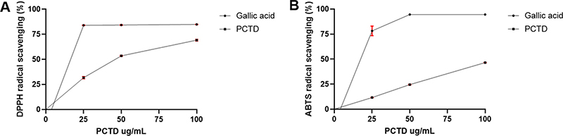
DPPH (A) and ABTS (B) radical-scavenging activities of PCTD.Gallic acid was used as a positive control. Values are presented as mean ± SD (n = 3).
2. Anti-inflammatory Effects
To assess potential cytotoxicity, cell viability was measured after treatment with different concentrations of PCTD. LPS stimulation increased the MTS signal compared with the untreated control. However, PCTD treatment at concentrations up to 200 ㎍/mL did not significantly reduce the MTS signal compared with the LPS-stimulated group (Fig. 2A).
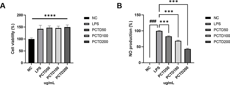
Cell viability (A) and nitrite levels (B) in LPS-stimulated RAW264.7 cells treated with PCTD.Values represent the mean ± SD (cell viability, n = 6; nitrite, n = 3). ###p < 0.001 vs. unstimulated cells; ****p < 0.0001, ***p < 0.001 vs. LPS-stimulated cells.
Nitric oxide (NO) is a reactive species produced during cellular inflammatory responses and is widely recognized as a representative indicator for evaluating anti-inflammatory activity. The LPS-treated group produced a large amount of nitrite compared with the control group. However, when extracts at concentrations of 50, 100, and 200 ㎍/mL were added under LPS-stimulated conditions, NO production exhibited a concentration-dependent inhibitory trend. Significant differences were observed between all treated groups and the LPS-treated group. Notably, at 200 ㎍/mL, PCTD reduced NO production by approximately 56.6% compared with the LPS-treated group (Fig. 2B).
PGE2 is an important inflammatory mediator associated with cyclooxygenase activity and inflammatory responses (Wallace, 2008; Halter, 2001). To evaluate the inhibitory effect of PCTD on LPS-induced macrophage inflammation, PGE2 levels were measured after treatment with different concentrations. The study demonstrated that PGE2 production in the LPS-treated group significantly increased compared to the control group. Compared with the LPS-treated group, PGE2 production was reduced by approximately 31.2% and 36.6% following treatment with 100 and 200 ㎍/mL PCTD, respectively. These results indicate that PCTD reduced LPS-induced PGE2 production (Fig. 3).
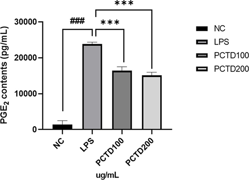
Effects of PCTD on prostaglandin E2 (PGE2) levels in the LPS-stimulated RAW264.7 cells.Values are presented as mean ± SD (n = 3). ###p < 0.001 vs. unstimulated cells; ***p < 0.001 vs. LPS-stimulated cells.
To further validate the anti-inflammatory properties of PCTD, pro-inflammatory cytokine levels in culture supernatants were measured via ELISA. LPS stimulation markedly increased the secretion of IL-6, IL-1β, G-CSF, and GM-CSF compared with the untreated control group.
PCTD treatment showed a decreasing trend in the levels of these cytokines, with a significant reduction in GM-CSF at 200 ㎍/mL. Specifically, GM-CSF secretion was reduced by approximately 17.3% compared with the LPS-treated group. These results indicate that PCTD attenuated LPS-induced inflammatory mediator production in RAW264.7 cells (Fig. 4).
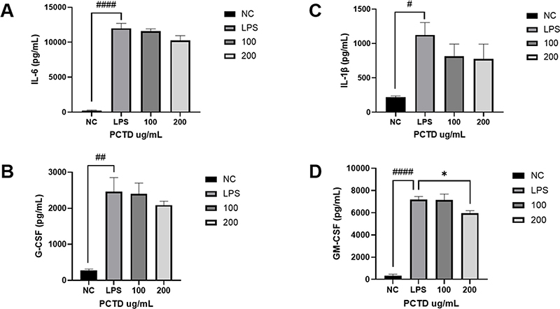
Effects of PCTD on the production of IL-6 (A), G-CSF (B), IL-1β (C), and GM-CSF (D) in RAW264.7 cells.Data are expressed as mean ± SD from three independent experiments (n = 3). ####p < 0.0001, ##p < 0.01, #p < 0.05 vs. unstimulated cells; *p < 0.05 vs. LPS-stimulated cells.
LPS stimulation significantly increased COX-2 and IL-1β expression, whereas COX-1 and IL-10 expression were significantly decreased compared with the normal control group. PCTD treatment significantly decreased COX-2 expression at 100 and 200 ㎍/mL, by approximately 41.7% and 57.1%, respectively, compared with the LPS-stimulated group, while IL-1β expression was significantly decreased at 200 ㎍/mL, by approximately 47.4%. In contrast, COX-1 and IL-10 expression were significantly increased by PCTD treatment at 200 ㎍/mL compared with the LPS-stimulated group, reaching approximately 2.54-fold and 3.75-fold of the LPS-treated levels, respectively (Fig. 5).
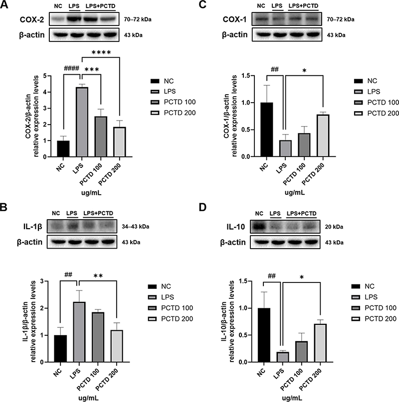
Effects of PCTD on COX-2 (A), IL-1β (B), COX-1 (C), and IL-10 (D) protein expression in LPS-stimulated RAW264.7 cells.Representative Western blot images and densitometric analyses are shown. Protein expression was normalized to β-actin. Data are expressed as mean ± SD from three independent experiments (n = 3). ####p < 0.0001, ##p < 0.01 vs. unstimulated cells; ****p < 0.0001, ***p < 0.001, **p < 0.01, *p < 0.05 vs. LPS-stimulated cells.
3. Gastric mucosal protective effects
To evaluate the protective effect of PCTD against acute gastric mucosal injury, an EtOH/HCl-induced acute gastric mucosal injury model was employed in mice. As shown in Fig. 6, the EtOH/HCl group exhibited a significantly higher gastric damage ratio than the normal control (NC) group. Compared with the EtOH/HCl group, the treatment group administered 50 ㎎/㎏ PCTD exhibited significantly reduced mucosal damage, with lesion area decreasing by approximately 13.70% to 6.61%, corresponding to a 51.7% reduction. The positive control group (R25) demonstrated a similar degree of improvement to the treatment group.
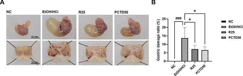
Effects of PCTD on EtOH/HCl-induced gastric mucosal injury in mice.Gross appearance (A) and gastric mucosal injury ratio (B). NC–Normal control group; EtOH/HCl–EtOH/HCl group; R25–EtOH/HCl-treated group administered ranitidine (25 ㎎/㎏); and PCTD50–EtOH/HCl-treated group administered PCTD (50 ㎎/㎏). ###p < 0.001 vs. NC group; *p < 0.05 vs. EtOH/HCl group.
H&E staining was performed to observe histopathological changes in the damaged gastric mucosa and those induced by PCTD administration. The EtOH/HCl group exhibited characteristic damage, including surface epithelial loss, mucosal folding, hemorrhage, and inflammatory cell infiltration. In the positive control group treated with 25 ㎎/㎏ ranitidine and the PCTD 50 ㎎/㎏ group, although epithelial damage, muscularis hemorrhage, and inflammatory cell infiltration were still visible, the histopathological changes appeared less pronounced compared with the EtOH/HCl group (Fig. 7).
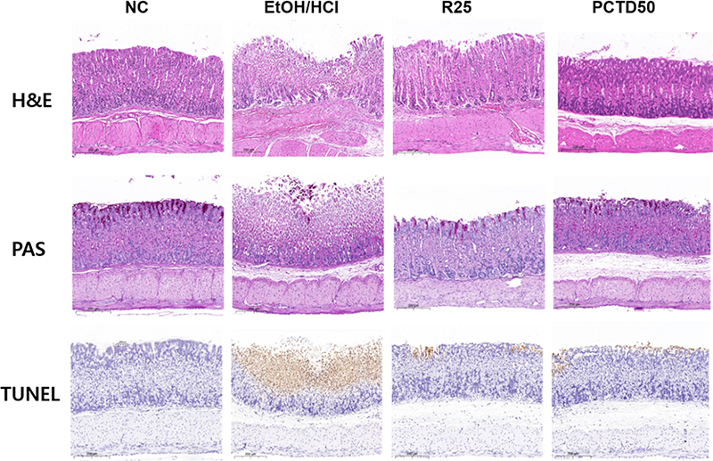
Effects of PCTD on histopathological changes in the stomach of mice with EtOH/HCl-induced gastric mucosal injury.Representative H&E-, PAS-, and TUNEL-stained gastric tissue sections are shown. NC–Normal control group; EtOH/HCl–EtOH/ HCl group; R25–EtOH/HCl-treated group administered ranitidine (25 ㎎/㎏); and PCTD50–EtOH/HCl-treated group administered PCTD (50 ㎎/㎏).
Analysis of mucus secretion function is an important indicator for evaluating gastric mucosal protective efficacy. Periodic acid-Schiff (PAS) staining enables visualization of mucus-secreting cells in the epithelial layer as purple-stained areas. PAS-positive staining appeared less extensive in the damaged areas than in the normal group. In contrast, representative sections from the PCTD-treated group showed more extensive PAS-positive staining than those from the EtOH/HCl group, with a staining pattern similar to that observed in the positive control group.
To assess DNA fragmentation associated with gastric mucosal injury, TUNEL staining was performed. Representative sections from the EtOH/HCl group showed more extensive TUNEL-positive staining than those from the normal group. In contrast, TUNEL-positive staining appeared less extensive in the ranitidine- and PCTD-treated groups than in the EtOH/HCl group.
DISCUSSION
Inflammation is a complex defensive physiological response triggered by the immune system to tissue injury, infection, or harmful stimuli (Zarrin et al., 2021). Gastritis is characterized by inflammatory damage to the gastric mucosa. Excessive apoptosis or necrosis of gastric epithelial cells can contribute to mucosal damage, leading to structural disruption and hemorrhage (Yan et al., 2024). Current therapeutic options for gastritis include antacids, proton pump inhibitors (PPIs), and histamine H2 receptor antagonists (Kim et al., 2025). Recently, pharmacologically active plant-derived compounds have received attention as promising therapeutic and preventive candidates for gastric mucosal injuries (Lee et al., 2022; Chen et al., 2025).
Ethanol and HCl aggravate gastric mucosal damage by inducing inflammatory responses, while hydrochloric acid further damages the gastric mucus layer and gastric tissue (Li et al., 2008). Therefore, to investigate the anti-inflammatory and gastric mucosal protective effects of PCTD, this study employed LPS-stimulated RAW264.7 macrophage inflammation models in vitro and an EtOH/HCl-induced acute gastric mucosal injury model in mice.
Oxidative stress is caused by an imbalance between oxidation and antioxidant defense systems. Excess ROS production can activate inflammatory pathways and promote the expression of inflammatory mediators and cytokines (Lingappan, 2018; Nam et al., 2021). Numerous studies have reported that plant extracts exhibit antioxidant activity through free radical scavenging (Hwang et al., 2023; Heckmann et al., 2024). In the present study, PCTD showed DPPH and ABTS radical scavenging activity, and the scavenging activity increased with PCTD concentration (Fig. 1). These results indicate that PCTD possesses antioxidant potential.
The anti-inflammatory efficacy of bioactive agents is commonly screened through LPS-induced macrophage models. LPS, a component of the outer membrane of Gram-negative bacteria, stimulates macrophages to release a range of inflammatory cytokines and mediators (Wang et al., 2017). Produced by inducible nitric oxide synthase (iNOS), NO serves as a key inflammatory mediator, with its overproduction exacerbating inflammatory pathways (Alderton et al., 2001). PCTD treatment did not significantly reduce the MTS signal at concentrations up to 200 ㎍/mL (Fig. 2A) and significantly reduced LPS-induced NO production (Fig. 2B).
PGE2 is another important inflammatory mediator associated with cyclooxygenase activity (Wallace, 2008), and regulation of excessive PGE2 production has been associated with the attenuation of inflammatory responses. Simultaneously, it drives the inflammatory cascade forward by stimulating immune cells to upregulate inflammatory cytokine production (Halter, 2001). IL-1β and IL-6 are representative pro-inflammatory cytokines involved in the amplification of inflammatory responses (Wang et al., 2017; Gong et al., 2021). Also, granulocyte colony-stimulating factor (G-CSF) and granulocyte-macrophage colony-stimulating factor (GM-CSF) are produced by macrophages during inflammatory responses and can contribute to the amplification of inflammation together with inflammatory cytokines (Lee and Park, 2015; Hwangbo et al., 2021). Recently, it has been reported that natural anti-inflammatory flavonoids such as quercetin not only inhibit major pro-inflammatory cytokines in stimulated macrophages but also downregulate growth factors such as G-CSF and GM-CSF (Kim and Park, 2016). In this study, PCTD significantly reduced LPS-induced PGE2 production (Fig. 3). Also, PCTD treatment showed decreasing trends in IL-6, IL-1β, and G-CSF levels, whereas GM-CSF was significantly reduced at 200 ㎍/mL (Fig. 4). These results support the anti-inflammatory activity of PCTD. COX-2 is associated with inflammatory prostaglandin production, whereas IL-10 plays an important role in limiting excessive inflammatory responses (DuBois et al., 1998; Saraiva et al., 2020). Similar changes in COX-2, PGE2, and IL-10 have been reported in LPS-stimulated RAW264.7 macrophages following anti-inflammatory treatment (Chou et al., 2003). Consistent with these findings, PCTD reduced COX-2 and IL-1β expression and increased COX-1 and IL-10 expression (Fig. 5).
Gastritis refers to inflammatory damage to the gastric mucosa caused by exogenous stimuli, often accompanied by features such as mucosal hyperemia, hemorrhage, and erosion (Kim et al., 2023). The gastric mucosal protective effect of PCTD was further evaluated in the EtOH/HCl-induced acute gastric mucosal injury model. EtOH/HCl administration caused extensive hemorrhagic lesions and mucosal damage, whereas PCTD treatment reduced the damaged gastric mucosal area (Fig. 6). These findings indicate that PCTD attenuated acute gastric mucosal injury induced by EtOH/HCl.
Histological findings further supported the protective effect of PCTD against EtOH/HCl-induced gastric mucosal injury. EtOH/HCl treatment caused epithelial disruption, hemorrhage, and inflammatory cell infiltration, whereas these histopathological changes were attenuated in the PCTD-treated group (Fig. 7). To protect the gastric mucosa from external stimuli, mucus-secreting cells in normal tissue secrete mucus. However, when stimulated, increased pepsin production and reduced mucosal blood flow can impair mucus secretion. Decreased mucus secretion weakens protection against external stimuli, leading to damage and inflammation in the mucosa, submucosa, and muscularis layers (Hagen, 2021; Jia et al., 2023). Previous studies have shown that ethanol-induced gastric injury is accompanied by disruption of the gastric epithelium and impairment of the mucus barrier (Li et al., 2021; Hagen, 2021). Consistent with these findings, representative PAS-stained sections showed less extensive positive staining in the EtOH/HCl group than in the normal group, whereas more extensive PAS-positive staining was observed following PCTD treatment (Fig. 7). In addition, to assess DNA fragmentation associated with gastric mucosal injury, TUNEL staining was performed. Representative TUNEL-stained sections showed more extensive positive staining in the EtOH/HCl group than in the normal control group, whereas staining appeared less extensive following PCTD treatment, with a pattern similar to that observed in the positive control group (Fig. 7). These qualitative observations suggest that PCTD treatment was associated with less pronounced gastric mucosal damage.
Because the present in vivo study evaluated a single PCTD dose, further studies using multiple doses would be useful to characterize the dose-response relationship. In addition, as ranitidine was used as the sole positive control, the present study could not distinguish acid-suppressive effects from direct mucosal-protective effects, which should be considered a limitation of the present study.
In this study, the antioxidant and anti-inflammatory activities of the hot-water extract of Phyllostachyos Caulis in Taeniam (PCTD) were confirmed through in vitro and in vivo assays. Together, these findings demonstrate that PCTD attenuates EtOH/HCl-induced acute gastric mucosal injury in mice. Further studies are needed to clarify the molecular mechanisms underlying these effects.
Acknowledgments
This work was supported by a grant from the Korea Institute of Oriental Medicine (Grant No. KSN2512030).
References
-
Alderton WK, Cooper CE and Knowles RG. (2001). Nitric oxide synthases: structure, function and inhibition. Biochemical Journal. 357:593-615.
[https://doi.org/10.1042/bj3570593]

-
An Y, Lee HW, Jung YC, Woo HJ, Kim YH, Kim HH, Jeong YJ, Abd Ei-Aty AM and Jeong JH. (2025). Phyllostachyos caulis in taeniam extract stimulates longitudinal bone growth via IGF-1 and JAK2/STAT5 signaling in rats. PLOS ONE. 20:e0331834.
[https://doi.org/10.1371/journal.pone.0331834]
-
Bibi Sadeer N, Montesano D, Albrizio S, Zengin G and Mahomoodally MF. (2020). The versatility of antioxidant assays in food science and safety—chemistry, applications, strengths, and limitations. Antioxidants. 9:709, https://www.mdpi.com/2076-3921/9/8/709, (cited by 2026 Oct 15).
[https://doi.org/10.3390/antiox9080709]
-
Chen D, Wang W, Chen X, Liang N, Li J, Ding W, Zhang H, Yang Z, Zhao H and Liu Z. (2025). Plant-derived extracts or compounds for Helicobacter-associated gastritis: A systematic review of their anti-helicobacter activity and anti-inflammatory effect in animal experiments. Chinese Medicine. 20:53.
[https://doi.org/10.1186/s13020-025-01093-2]
-
Chou TC, Fu E and Shen EC. (2003). Chitosan inhibits prostaglandin E2 formation and cyclooxygenase-2 induction in lipopolysaccharide-treated RAW 264.7 macrophages. Biochemical and Biophysical Research Communications. 308:403-407.
[https://doi.org/10.1016/S0006-291X(03)01407-4]
-
DuBois RN, Abramson SB, Crofford L, Gupta RA, Simon LS, Van De Putte LB and Lipsky PE. (1998). Cyclooxygenase in biology and disease. The FASEB Journal. 12:1063-1073.
[https://doi.org/10.1096/fasebj.12.12.1063]
-
Gong G, Zhao R, Zhu Y, Yu J, Wei B, Xu Y, Cui Z and Liang G. (2021). Gastroprotective effect of cirsilineol against hydrochloric acid/ethanol-induced gastric ulcer in rats. The Korean Journal of Physiology & Pharmacology. 25:403-411.
[https://doi.org/10.4196/kjpp.2021.25.5.403]
-
Hagen SJ. (2021). Mucosal defense: Gastroduodenal injury and repair mechanisms. Current Opinion in Gastroenterology. 37:609-614.
[https://doi.org/10.1097/MOG.0000000000000775]
-
Halter F. (2001). Cyclooxygenase 2—implications on maintenance of gastric mucosal integrity and ulcer healing: Controversial issues and perspectives. Gut. 49:443-453.
[https://doi.org/10.1136/gut.49.3.443]
-
Heckmann M, Stadlbauer V, Drotarova I, Gramatte T, Feichtinger M, Arnaut V, Atzmüller S, Schwarzinger B, Röhrl C, Blank-Landeshammer B and Weghuber J. (2024). Identification of oxidative-stress-reducing plant extracts from a novel extract library—comparative analysis of cell-free and cell-based in vitro assays to quantitate antioxidant activity. Antioxidants. 13:297.
[https://doi.org/10.3390/antiox13030297]
-
Hwang J, Jang B, Choi YW, Han IO and Oh ES. (2023). Synergistic anti-inflammatory effects of ethanol extracts from Chrysanthemum zawadskii flower and Cudrania tricuspidata fruit occur via inhibition of the NF-κB signaling pathway. Evidence-Based Complementary and Alternative Medicine. 2023:8198228.
[https://doi.org/10.1155/2023/8198228]
-
Hwangbo H, Ji SY, Kim MY, Kim SY, Lee H, Kim GY, Kim S, Cheong J and Choi YH. (2021). Anti-inflammatory effect of auranofin on palmitic acid and LPS-induced inflammatory response by modulating TLR4 and NOX4-mediated NF-kappa-B signaling pathway in RAW264.7 macrophages. International Journal of Molecular Sciences. 22:5920.
[https://doi.org/10.3390/ijms22115920]
-
Jia X, He Y, Li L and Xu D. (2023). Pharmacological targeting of gastric mucosal barrier with traditional Chinese medications for repairing gastric mucosal injury. Frontiers in Pharmacology. 14:1091530.
[https://doi.org/10.3389/fphar.2023.1091530]
-
Jin GH, Park SY, Kim EY, Ryu EY, Kim YH, Park GT and Lee SJ. (2012). Anti-inflammatory activity of bambusae caulis in taeniam through heme oxygenase-1 expression via Nrf-2 and p38 MAPK signaling in macrophages. Environmental Toxicology and Pharmacology. 34:315–323.
[https://doi.org/10.1016/j.etap.2012.05.001]
-
Kim A, Im M, Yim NH, Jung YP and Ma JY. (2013). Aqueous extract of bambusae caulis in taeniam inhibits PMA-induced tumor cell invasion and pulmonary metastasis: Suppression of NF-κB activation through ROS signaling. PLOS ONE. 8:e78061.
[https://doi.org/10.1371/journal.pone.0078061]
-
Kim BK, Huh J, Kim SG, Ahn JY and Kim JW. (2025). Pharmacological treatment of gastritis: A narrative review with a systematic literature search. Gut and Liver. 19:783-794.
[https://doi.org/10.5009/gnl250267]
-
Kim GH, Choi MG, Kim JI, Lee ST, Chun HJ, Lee KL, Choi SC, Jang JY, Lee YC, Kim JG, Kim KB, Shim KN, Sohn CI, Kim SK, Kim SG, Jang JS, Kim NY, Jung HY, Park HJ, Huh KC, Lee KJ, Hong SJ, Baek S, Han JJ and Lee OY. (2023). Efficacy and safety of fexuprazan in patients with acute or chronic gastritis. Gut and Liver. 17:884–893.
[https://doi.org/10.5009/gnl220457]
-
Kim JH, Chun SY, Ohk SO, Kim SH, Kim JW, Lee SO, Kim HG and Kim SJ. (2021). Amelioration of alcohol-induced gastric mucosa damage by oral administration of food-polydeoxyribonucleotides. Molecular Medicine Reports. 24:790.
[https://doi.org/10.3892/mmr.2021.12430]
-
Kim YJ and Park W. (2016). Anti-inflammatory effect of quercetin on RAW 264.7 mouse macrophages induced with polyinosinic-polycytidylic acid. Molecules. 21:450. https://www.mdpi.com/1420-3049/21/4/450, (cited by 2026 Oct 15).
[https://doi.org/10.3390/molecules21040450]
-
Lee J and Park W. (2015). Anti-inflammatory effect of wogonin on RAW 264.7 mouse macrophages induced with polyinosinic-polycytidylic acid. Molecules. 20:6888-6900. https://www.mdpi.com/1420-3049/20/4/6888, (cited by 2026 Oct 15).
[https://doi.org/10.3390/molecules20046888]
-
Lee JH, Kwak HJ, Shin DC, Seo HJ, Park SJ, Hong BH, Shin MS, Kim SH and Kang KS. (2022). Mitigation of gastric damage using Cinnamomum cassia extract: Network pharmacological analysis of active compounds and protection effects in rats. Plants. 11:716.
[https://doi.org/10.3390/plants11060716]
-
Li CY, Xu HD, Zhao BT, Chang HI and Rhee HI. (2008). Gastroprotective effect of cyanidin 3-glucoside on ethanol-induced gastric lesions in rats. Alcohol. 42:683-687.
[https://doi.org/10.1016/j.alcohol.2008.08.009]
-
Li WS, Lin SH, Chu CH, Chang YK, Zhang X, Lin CC and Tung YT. (2021). The gastroprotective effect of naringenin against ethanol-induced gastric mucosal injury in mice through regulating inflammation and apoptosis. International Journal of Molecular Sciences. 22:11985.
[https://doi.org/10.3390/ijms222111985]
-
Lim D, Cho Y, Kim W, Jeong S, Jang YP and Kim J. (2017). Extract of bambusae caulis in taeniam inhibits cigarette smoke-induced pulmonary and intestinal inflammation. Experimental Biology and Medicine. 242:102-112.
[https://doi.org/10.1177/1535370216664429]
-
Lingappan K. (2018). NF-κB in oxidative stress. Current Opinion in Toxicology. 7:81-86.
[https://doi.org/10.1016/j.cotox.2017.11.002]
- Lu B, Liu L, Zhen X, Wu X and Zhang Y. (2010). Anti-tumor activity of triterpenoid-rich extract from bamboo shavings (caulis bamfusae in taeniam). African Journal of Biotechnology. 9:6430-6436.
-
Nam HH, Nan L and Choo BK. (2021). Inhibitory effects of Camellia japonica on cell inflammation and acute rat reflux esophagitis. Chinese Medicine. 16:6.
[https://doi.org/10.1186/s13020-020-00411-0]
-
Oh TY, Ahn GJ, Choi SM, Ahn BO and Kim WB. (2005). Increased susceptibility of ethanol-treated gastric mucosa to naproxen and its inhibition by DA-9601, an Artemisia asiatica extract. World Journal of Gastroenterology. 11:7450-7456.
[https://doi.org/10.3748/wjg.v11.i47.7450]
-
Ra J, Lee S, Kim HJ, Jang YP, Ahn H and Kim J. (2010). Bambusae Caulis in Taeniam extract reduces ovalbumin-induced airway inflammation and T helper 2 responses in mice. Journal of Ethnopharmacology. 128:241–247.
[https://doi.org/10.1016/j.jep.2010.01.023]
-
Saraiva M, Vieira P and O'Garra A. (2020). Biology and therapeutic potential of interleukin-10. Journal of Experimental Medicine. 217:e20190418.
[https://doi.org/10.1084/jem.20190418]
-
Wallace JL. (2008). Prostaglandins, NSAIDs, and gastric mucosal protection: Why doesn’t the stomach digest itself? Physiological Reviews. 88:1547-1565.
[https://doi.org/10.1152/physrev.00004.2008]
-
Wang S, Chen M, Xu S, Zhang J, Wang Z, Ma X, Yang X and Liu C. (2017). Effect of luteolin on inflammatory responses in RAW264.7 macrophages activated with LPS and IFN-γ. Journal of Functional Foods. 32:123–130.
[https://doi.org/10.1016/j.jff.2017.02.018]
-
Yan S, Bao S, Chen T, Chen J, Zhang J, Hu X, Liang Y, Zhou X and Li J. (2024). Cinnamaldehyde alleviates aspirin-induced gastric mucosal injury by regulating PI3K/AKT pathway-mediated apoptosis, autophagy and ferroptosis. Phytomedicine. 132:155791.
[https://doi.org/10.1016/j.phymed.2024.155791]
-
Yang YY, Li KM, Xu GF, Wang CD, Xiong H, Wang XZ, Wang CH, Zhang BY, Jiang HX, Sun J, Xu Y, Zhang LJ, Zheng HX, Xing XB, Wang LJ, Zuo XL, Ding SG, Lin R, Chen CX, Wang XW and Li JN. (2024). Clinical manifestation, lifestyle, and treatment patterns of chronic erosive gastritis: A multicenter real-world study in China. World Journal of Gastroenterology. 30:1108-1120.
[https://doi.org/10.3748/wjg.v30.i9.1108]
-
Zarrin AA, Bao K, Lupardus P and Vucic D. (2021). Kinase inhibition in autoimmunity and inflammation. Nature Reviews Drug Discovery. 20:39-63.
[https://doi.org/10.1038/s41573-020-0082-8]